Pinched Nerve

(Radiculopathy)

Our Awards

Your central nervous system (CNS) stays busy around the clock, transmitting information to and from the brain and body to produce movement, detect environmental changes and signal when something is not quite right. Sometimes your nerves can become compressed or “pinched” by other structures in your body, including bones, muscles, tendons and ligaments. Even your spinal discs can pinch your nerves if they become inflamed, or herniated.

Your spine provides a protective tunnel for your CNS as it descends from your brain and branches out to innervate various parts of your body. Nerve roots emerge from between your vertebrae along your spinal column, to act on specific areas of your body. When a nerve becomes pinched, it can cause pain anywhere along its pathway. For example, a pinched sciatic nerve in your low back may cause pain that shoots down your leg, all the way to your foot.

Radiculopathy can stem from one of three areas of the spine:
bg
Cervical radiculopathy

occurs from pressure on a nerve root in your neck. Symptoms may include weakness, burning or tingling, or numbness in your shoulders, arms, hands, or fingers.

bg
Thoracic radiculopathy

is relatively uncommon, and is caused by a compressed nerve in the upper back portion of your spine, creating pain in your chest and torso.

bg
Lumbar radiculopathy

arises from pressure on a nerve root in your low back. It may cause hip pain or sciatic nerve pain that shoots down your leg. It can also contribute to incontinence, sexual dysfunction, or even paralysis in severe cases.

Pinched Nerve Symptoms and Causes include:

  • Poor postural habits
  • Trauma
  • Stress
  • Overuse or misuse of body parts
  • Tight or weakened muscles
  • Sedentary lifestyle
  • Overweight or obese
  • Poor physical condition
  • Wear and tear from aging

Diagnosis of Radiculopathy

Diagnosis of radiculopathy begins with a review of patient history and a physical exam. An X-ray, ultrasound or MRI scan may be used to rule out fractures or other sources of pain, and to confirm the diagnosis. Other diagnostic tests may include a nerve conduction study to identified a damaged nerve, and electromyography (EMG) to determine if there is damage to the nerves leading to a muscle.

bg

Pinched Nerve Treatment Options

Treatment of radiculopathy is geared to reducing pain and relieving pressure on nerve roots. Some common treatment measures include:

Rest and activity change:

Reducing your physical activities and refraining from activities that cause pain may help ease inflammation and pressure on your nerves.

Splinting or bracing:

You may be given a split or brace to immobilize the painful area.

Medications:

NSAIDs like ibuprofen or naproxen can help reduce pain and inflammation. Muscle relaxers can help ease compression caused by tight or spastic muscles.

Corticosteroid injections:

Steroids can sometimes help relieve pain and reduce inflammation.

Physical Therapy:

Certain exercises and stretches combined with lifestyle modification can help relieve nerve pressure and prevent its return.

Surgery:

When conservative treatment is unsuccessful in relieving radiculopathy, surgical intervention may be a last resort to remove bone spurs or a portion of a herniated disk in the spine.

Clinical Case Studies
NYDNRehab


Peroneal Nerve Entrapment

Our female patient came to us after unsuccessful treatment at another clinic. She presented with painful hip snapping, leg paresthesia (tingling/numbness), low back pain, sacroiliac(SI) joint pain, and gluteal pain.

learn more

atient with Facial Pain and Complex Neurological Dysfunction Successfully Treated with Alternative Therapies

Our patient, a 28 year-old male, had been suffering for over two years with unexplained facial and upper body pain. He had seen two separate neurologists and undergone MRIs of his brain and cervical spine, with no concrete diagnosis.

learn more
bg
bg
bg

Pinched Nerve Doctor at NYDNRehab

The nerve pain specialists at NYDNRehab take a holistic approach to treating radiculopathy. We go beyond merely treating your symptoms, pulling out all the stops to get to the source of your pain and eliminate it. Using cutting-edge technology and innovative treatment methods, we correct misalignmentsm imbalances and motor deficiencies to resolve radiculopathy at its source.

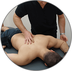
Treatment approaches for radiculopathy at NYDNRehab include: Dynamic Neuromuscular Stabilization (DNS):

This safe and effective treatment method for back, neck, and shoulder pain restores dysfunction of the locomotor system to enable nerves to move freely within your body.

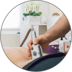
Extracorporeal shockwave therapy (ESWT):

By sending shock waves to the site of injury, ESWT helps the body heal itself, accelerating cell regeneration while relieving inflammation.

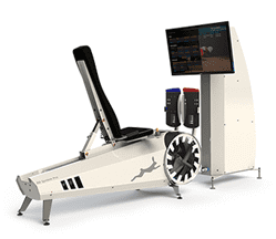
Computer Assisted Rehabilitation Environment (C.A.R.E.N):

This fully immersive virtual reality environment can help retrain your body to move more efficiently, relieving nerve pressure and restoring optimal functional movement.

Chiropractic manipulation:

Manipulating the spine can relieve nerve pressure and help restore correct spinal alignment.

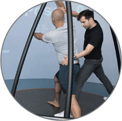
Posture education and retraining:

Learning to properly align your body during sitting, standing and walking can help to relieve radiculopathy and prevent its return.

Walking and running gait retraining:

Mechanical deficiencies in the way you walk and run often contribute to radiculopathy. They can be corrected to relieve pain and prevent recurring incidents.

Exercises and stretches to restore muscle balance:

Tight, lax or weak muscles can contribute to radiculopathy. Exercise can restore muscle tone to relieve pressure on nerve roots. You do not have to live with pinched nerve pain. The nerve pain specialists at NYDNRehab are dedicated to identifying the source of your pain and eliminating it for good.

Research at NYDNRehab

bg

In this instance, an athlete was originally diagnosed with minor quadriceps muscle strain and was treated for four weeks, with unsatisfactory results. When he came to our clinic, the muscle was not healing, and the patients’ muscle tissue had already begun to atrophy.

Upon examination using MSUS, we discovered that he had a full muscle thickness tear that had been overlooked by his previous provider. To mitigate damage and promote healing, surgery should have been performed immediately after the injury occurred. Because of misdiagnosis and inappropriate treatment, the patient now has permanent damage that cannot be corrected.

The most important advantage of Ultrasound over MRI imaging is its ability to zero in on the symptomatic region and obtain imaging, with active participation and feedback from the patient. Using dynamic MSUS, we can see what happens when patients contract their muscles, something that cannot be done with MRI. From a diagnostic perspective, this interaction is invaluable.

Dynamic ultrasonography examination demonstrating
the full thickness tear and already occurring muscle atrophy
due to misdiagnosis and not referring the patient
to proper diagnostic workup

Demonstration of how very small muscle defect is made and revealed
to be a complete tear with muscle contraction
under diagnostic sonography (not possible with MRI)

image

Complete tear of rectus femoris
with large hematoma (blood)

image

Separation of muscle ends due to tear elicited
on dynamic sonography examination

bg
Buy now 3D Gait
Payment Success
Request Telehealth Request Telehealth Request in office visit Book now