Biofeedback Motor Control Training And Analysis

Biomechanical Analysis With Real Time Force Plate Technology

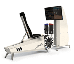
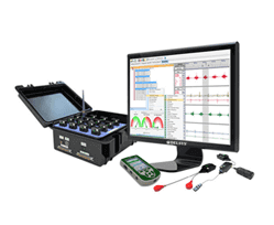
Biomechanical analysis can register the forces involved with athletic movement. When these forces exceed normal there is excessive stress to joints, muscles and ligaments. The excessive forces are always the result of poor muscle coordination, improper technique (motor control) and muscular weakness. The in depth biomechanical analysis allows differentiation of these factors and provides proper strategy to reduce injury and improve performance. Read more…

Biofeedback Motor Control Training

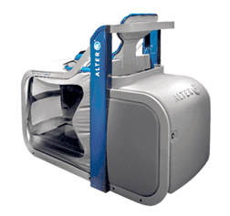
We’ve made it our mission to help our patients find the pathway to recovery as quickly as possible, providing them with a better quality of life and improved functioning. Biofeedback motor control training offers a groundbreaking approach that has had promising results for our patients with lower extremity disorders. If you are the victim of a host of injuries involving the knees or hips due to an active lifestyle, biofeedback motor control could be the answer you are looking for as you journey the road to rehabilitation. Read more…

bg

In this instance, an athlete was originally diagnosed with minor quadriceps muscle strain and was treated for four weeks, with unsatisfactory results. When he came to our clinic, the muscle was not healing, and the patients’ muscle tissue had already begun to atrophy.

Upon examination using MSUS, we discovered that he had a full muscle thickness tear that had been overlooked by his previous provider. To mitigate damage and promote healing, surgery should have been performed immediately after the injury occurred. Because of misdiagnosis and inappropriate treatment, the patient now has permanent damage that cannot be corrected.

The most important advantage of Ultrasound over MRI imaging is its ability to zero in on the symptomatic region and obtain imaging, with active participation and feedback from the patient. Using dynamic MSUS, we can see what happens when patients contract their muscles, something that cannot be done with MRI. From a diagnostic perspective, this interaction is invaluable.

Dynamic ultrasonography examination demonstrating
the full thickness tear and already occurring muscle atrophy
due to misdiagnosis and not referring the patient
to proper diagnostic workup

Demonstration of how very small muscle defect is made and revealed
to be a complete tear with muscle contraction
under diagnostic sonography (not possible with MRI)

image

Complete tear of rectus femoris
with large hematoma (blood)

image

Separation of muscle ends due to tear elicited
on dynamic sonography examination

bg
Buy now 3D Gait
Payment Success
Request Telehealth Request Telehealth Request in office visit Book now