Knee Pain Treatment
Knee pain is one of the most common physical injuries and has many possible causes. For this reason, it can be difficult but crucial to get an accurate diagnosis for treatment of knee pain. Patients may develop knee pain as a result of arthritis, ligament injuries, cartilage injuries, dislocated kneecap, bursitis, or patellar tendinitis. Luckily there are a variety of knee pain treatment options.
Anatomy of the Knee
The knee is a complex joint made up of three different compartments, each with its own unique functions and structures.
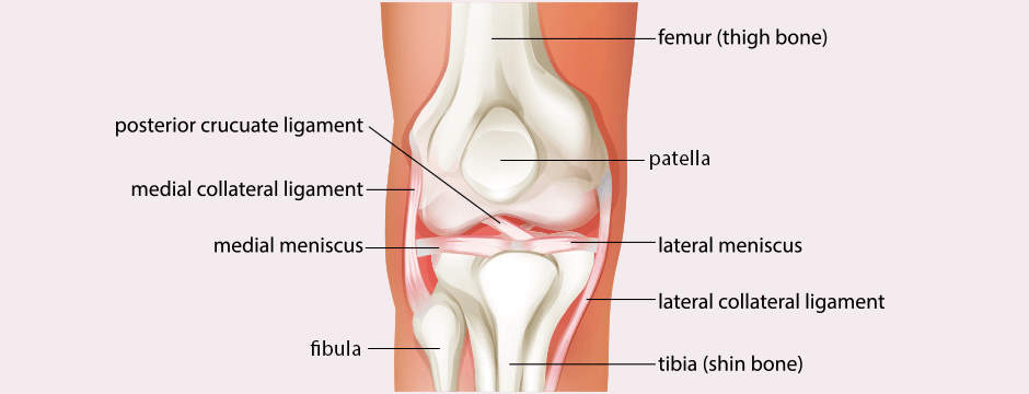
The inner (medial) compartment and the outer (lateral) compartments of the knee are formed by the junction of the femur and the shinbone. Where the kneecap (patella) and the front part of the femur meet, a third compartment is created, called the patellofemoral joint. The first two compartments allow the patient to walk on flat terrain, while the third compartment assists in activities like walking on uneven surfaces, going up and down stairs, kneeling, and standing up.
Below the kneecap the large patellar tendon attaches to the front of the tibia. Large blood vessels pass behind the knee, in the area known as the popliteal space. The knee is surrounded on the front side by the quadriceps muscles and on the back side by the hamstrings, which work together to flex and extend the knee joints.
A joint capsule surrounds the knee joint, held together by ligaments. The medial collateral ligament (MCL) and lateral collateral ligament (LCL) provide the knee with strength and stability. The MCL attaches the medial side of the femur to the medial side of the tibia, while the LCL attaches the lateral side of the femur to the lateral side of the fibula. Together the MCL and LCL prevent the femur from sliding around, while the anterior cruciate ligament (ACL) prevents it from sliding backward on the tibia. The posterior cruciate ligament (PCL) is the strongest knee ligament, and prevents the femur from sliding forward on the tibia.
The meniscus is a thickened pad of cartilage between the knee bones that creates a smooth surface for motion and absorbs pressure on the knee when the body is upright. The medial and lateral menisci, two C-shaped pieces of cartilage, lie between the femur and tibia and act as shock absorbers for the rest of the lower body.
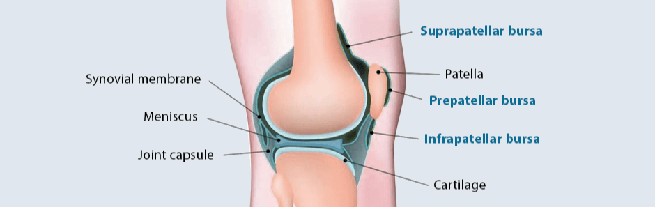
Around the knee joint lie bursae, small, cushiony sacs filled with fluid that rest between bones and surrounding tissue. When the patient is moving, bursae lubricate soft tissue and offer a gliding surface to reduce the friction of the tendons. Notably, the prepatellar bursa rests atop the kneecap, while the anserine bursa lurks within the knee, beneath the knee joint.
Causes of knee pain
Causes of the knee pain can be divided into multiple categories:
- Anterior knee pain due to patello-femoral and extensor mechanism causes
- Anterior knee pain due to knee joint issues
- Medial knee pain
- Lateral knee pain
- Posterior knee pain
Symptoms of knee pain
Knee arthritis is a condition typically affecting two or more compartments in the knee, though in rare cases it may be confined to the patellofemoral compartment. Symptoms of knee arthritis may include pain in the front part of the knee behind the patella, that is exacerbated by walking on inclined terrain, going up and down stairs, and standing up. However, the condition may pass unnoticed when the patient is walking on level ground.
Women are more likely to experience knee arthritis than men, and to require treatment for knee pain. Patients are more susceptible to this condition if they have excessive hip anteversion, a condition in which the neck of the femur rotates too far in the hip socket and pulls sideways on the patella. It is] also more common in patients suffering from patellofemoral dysplasia, in which one of the trochlea grooves is so misshapen that it no longer matches the patellar surface, causing the cartilage to deteriorate.
In certain cases requiring chronic knee pain treatment, patients with these conditions can suffer episodes of total patella dislocation. Also known as patellar instability, this condition causes repeated episodes of damage to the cartilage coating on the patella and predisposes patients to early patellofemoral arthritis. Moreover, because patellofemoral arthritis usually affects both legs, patients undergoing knee joint pain treatment may need to be examined in both, even if they are only experiencing symptoms in one knee.
Treatment for medial knee pain
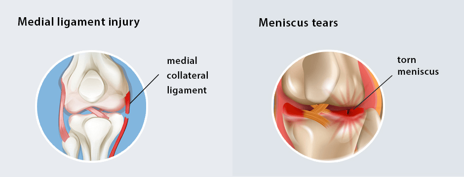
Because the medial patellofemoral ligament aids the knee in providing sideways movement and balance, medial patellofemoral ligament injury is a serious condition requiring medial knee pain treatment. The injury may take the form of a slight strain, a slight tear, or a total rupture necessitating surgery. A physician may also need to treat the surrounding area, because injury to the medial ligament typically spreads to the rest of the knee and may damage the medial meniscus, the cartilage, and the quadriceps.
The onset of medial ligament injury is normally accompanied by a sudden, jerky movement, like a twisting landing, in which the knee collapses in on itself. At times the injured party will feel a palpable tear in the knee or hear an audible popping sound in the area of damage. Pain will be accompanied by swelling, followed by constant throbbing, soreness, and limping.
Meniscus tears are usually the result of traumatic injury (commonly reported by athletes) and degeneration (common in the elderly). The meniscus may be torn when the knee joint is bent and the knee becomes twisted. Medial knee pain treatment then becomes necessary, especially if the injury is accompanied by years of overload to the medial knee joint.
These three medial knee problems together are known as the “unhappy triad,” such as happens to athletes when struck on the inside of the knee. Symptoms of meniscus tears may include pain, swelling, and joint locking, or the inability to straighten the joint, which occurs when a piece of the torn meniscus physically impinges inside the knee joint’s surfaces.
Knee Swelling and Pain Treatment
Because in its early stages patellofemoral arthritis is relatively benign, patients may walk on level ground for miles without experiencing pain or disability. Consequently the condition may be far advanced before the patient seeks inner knee pain treatment.
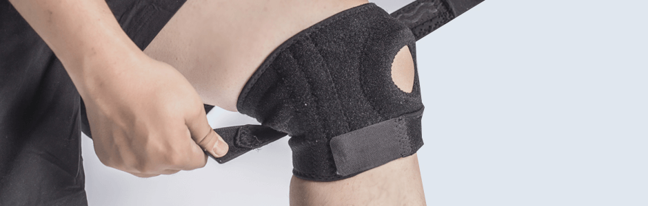
Depending on the degree of cartilage damage, treatment for knee pain may include measures such as wearing a brace. A physician may also recommend physical therapy as a form of inner knee pain treatment that strengthens and stretches the quadriceps muscles while improving patella tracking.
Victims of knee arthritis or related knee-pain injuries may experience relief by taking non-steroidal anti-inflammatory drugs (NSAIDs) such as ibuprofen. However, these should be taken only sparingly and with the approval and supervision of a physician. Knee pain treatment injections such as intra-articular steroid injections, or cortisone shots, are an alternative form of knee joint pain treatment and may be used during the acute phase to reduce pain and intra-articular inflammation. Knee pain treatment injections are another form of treatment for arthritic knees.This type of injection lubricates the joint and allows rehabilitation to progress.
Conventional methods of knee pain treatment are generally helpful for struggling patients, especially those who need to climb stairs. However, as the knee continues to degenerate, increased bone loss may result in chronic knee pain. Treatment in this case should target unloading of the knee.
In the case of a torn meniscus, effective knee pain treatments may include cryotherapy and immobilization to lessen swelling and pain in the joint. A physician may recommend an MRI evaluation to reveal abnormalities in the meniscus and to determine whether surgery is necessary.
Rehab and physical therapy versus regenirative injections
Regenerative injections may be needed in the later stages of knee degeneration, but in the early stagees physical therapy plays a key role. The knee joint is totally dependent on the movement of the hip and foot joints, and therefore normalizing movement in those joints is key. Therapy may include retraining gait mechanics, improving muscle strength and flexibility, and enhancing coordination and motor control in the early stages of any knee issues.
Knee Pain Treatment at NYDNRehab
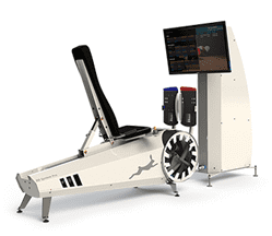
At NYDNRehab we specialize in various types of knee pain treatment, including chronic knee pain treatment, inner knee pain treatment, and anterior knee pain treatment. Our biofeedback motor control training uses real-time force plate analysis to treat injuries and conditions of the lower extremities. This training has been successfully used for treatment and post-surgical rehabilitation, and has proven extremely effective in treating injuries procured in prolonged, high-exertion physical activities like figure skating, tennis, running, and ballet.
We employ the most advanced and comprehensive treatment of meniscus tears, ACL/PCL tears, patellofemoral syndrome, and various forms of bursitis, providing diagnosis and physical therapy for anterior knee pain treatment and chronic knee pain treatment.
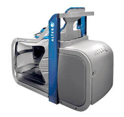
For those suffering from degeneration of knee tendons, in the tendons or who have encountered tension between the hamstrings and quadriceps, extracorporeal shockwave therapy (ESWT) is an inside knee pain treatment designed to relieve pressure and increase blood circulation to the injured areas. C.A.R.E.N, our computer assisted rehabilitation environment, is a fully immersive environment that uses principles of virtual reality and feedback training to restore walking and weight bearing.
TeleHealth at NYDNRehab
Get all the benefits of physical therapy and chiropractic care via safe and effective online sessions, all from the privacy and convenience of your home, office or hotel room. Never miss a session, and keep moving toward your physical therapy goals.