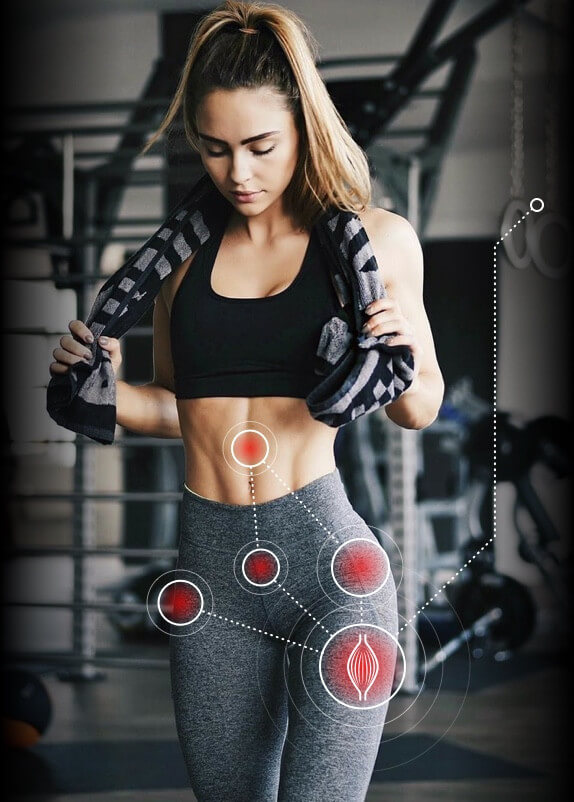
The hip is a prominence of the pelvis extending from the waist to the upper legs and is one of the most central and stabilizing locations in the body. Because of its importance to the workings of the lower extremities, the hip is prone to a greater number of injuries than many other parts of the body. Athletes and runners, especially, are especially susceptible to hip injuries. Hip injury treatment is often necessary for individuals suffering conditions like pinched nerve, labral tears, stress fracture, hip bursitis, impingement, and strained hip flexor.
Hip flexor strain refers to a condition affecting the muscles that allow flexion in the hips. Prominent among them are the iliopsoas muscles (the psoas major and iliacus muscle) and the muscles in the interior compartment of the thigh (the rector femoris and sartorius). The most important of these, the iliopsoas, is pivotal for standing, running, and walking. Strained hip flexor treatment includes conventional treatments like icing, resting, and stretching. Stretching of the hip flexors and quadriceps muscles and improve strength and flexibility.
The iliopsoas is sometimes injuried when the thigh is forced to extend or is blocked during active extension. This can occur, for example, in soccer players who are struck on the leg as their leg is extending to kick the soccer ball. It can also occur during weight lifting or uphill running.
Pubic symphysis dysfunction is a major cause of groin pain in some patients. This is a condition affecting a joint cartilage located between the left and right pubic bones above the genitalia and anterior to the bladder. Typically pubic symphysis dysfunction is the result of injury during high-speed cutting activity. It may also concurrently with adductor strain. In rare cases widening of the pelvic joints during pregnancy can harm the joint cartilage, resulting in dysfunction. Symptoms may include tenderness and swelling in the groin area and difficulty getting out of chairs, walking, going upstairs, and climbing into bed. Sometimes the pain is accompanied by a clicking sensation.
Stress fractures are another common source of hip pain in athletes—both femoral stress neck fractures and pubic ramus stress fractures. The pubic ramis is located at the front side of the pelvis and is divided into the inferior and superior pubic ramis, while the femoral neck is a flattened region near the top of the femur (thigh bone).
Pubic ramis stress fractures account for a small number of athletic stress fractures. They are especially common, however, in long-distance runners who undergo sudden changes in speed or intensity. Symptoms will include a gradual feeling of pain that is exacerbated by physical exercise.
Femoral neck stress fractures also affect long-distance runners and can result not only from dramatic changes in routine but also from overuse, impaired bone metabolism, and muscle fatigue resulting in a loss of shock absorption. Risk factors can be exacerbated by training errors, improper footwear, and running on uneven surfaces. Symptoms may include pain in the groin, hip, or thigh that that lessens when the patient is not active.
Pubic ramus hip stress fracture treatment consists of avoiding activities that cause pain for between four to six weeks. While resting and recovering, the patient should engage in non-weight-bearing activities and exercises that stretch the adductor muscle group, with the ultimate goal of gradually transitioning back into normal activity. Within three to five months most athletes will show signs of recovery. Treatment for femoral neck stress fractures varies according to the nature of the injury. Compression fractures, which are common in younger patients, can normally be treated with conventional treatments if the fracture involves less than half the width of the femoral neck. A doctor will recommend avoiding weight-bearing activities, and in more extreme cases may prescribe a short bed rest. He or she will then gradually supervise a return to normal activities.
The hip has a labrum, a ring of articular cartilage, that deepens the acetabulum, a vast depression in the pelvis. Hip labral tears generally occur following some form of trauma, such as slipping or dislocation, although they are also sometimes associated with osteoarthritis. Symptoms of hip labral tear include pain during twists or pivots and pain during extension. These may be accompanied by a loud clicking sound and a feeling that the hip is “giving way.” Treatment for hip labral tear includes conservative management with physical therapy and anti-inflammatory medication.
Hip impingement, or femoroacetabular impingement (FAI) is a condition in which the bones of the hip are abnormally shaped and don’t fit together. As a result, the bones brush up against each other and cause friction. Although recognition of hip impingement is difficult, proper hip impingement treatment is important because the disorder may lead to osteoarthritis in young adults with otherwise normal anatomies. Symptoms of FAI include groin pain, pain over the greater trochanter, and grinding or popping sounds. Patients may also have trouble flexing and rotating, and may experience pain after periods of prolonged sitting.
Hip bursitis is an affliction of the bursae, small sac-like cavities that help lubricate the soft tissues around the joints and alleviate the friction that results when bones rub against each other. While bursae exist wherever there are joints in the body, the most commonly injured bursae in the hip area are the ischial, iliopsoas, and greater trochanteric bursitis. Typically this condition results from inflammation as a result of excessive friction and traumatic injury. Iliopsoas bursitis often occurs in athletes who are required to use their hip flexors excessively (soccer, ballet, hurdling), while greater trochanteric bursitis is often a result of overuse. Symptoms include pain when running, climbing stair, or standing for long periods of time. Treatment for hip bursitis is generally non-surgical and consists of rest, anti-inflammatory medication, and exercises for stretching. If the condition proves resist to conservative treatment, a physician may recommend corticosteroid injection to lessen inflammation and pain.
Meralgia paresthetica, or lateral femoral cutaneous nerve entrapment, is a condition that causes pinching of a nerve in the femur. While initially this condition was thought to proceed surgical treatments like appendectomy and bone grafting, it has more recently been reported in diabetic and obese patients, and in those wearing tight clothing on the lower half of their bodies. Athletes suffering from meralgia paresthetica may feel pain, numbness, or tingling above the thigh. Prolonged flexion or an increase in muscle mass can also be factors. As is typical for treatment of pinched nerve in hip, lateral femoral entrapment is generally treatable with non-surgical treatments like heat, physical therapy, and non-steroidal anti-inflammatory medications.
Hip tendinitis is a broad term used to describe a condition of inflammation or degeneration of tendons in the hips. The patient will notice a gradual increase in pain over time, along with tenderness at the site of the affected area. He or she may encounter discomfort when stretching adjoining muscles, and stiffness when first getting out of bed. Tendinitis hip treatment includes rest, gentle stretching, and, once the pain has subsided, a program of strengthening exercises for the injured muscles.
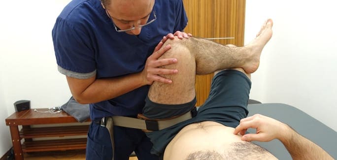
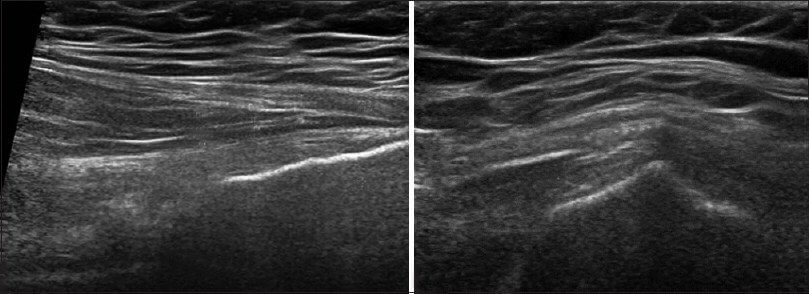
Patients needing treatment for groin pain or hip strain treatment will find comprehensive treatment at the New York DNR. We use musculoskeletal ultrasonography to analyze the integrity of the tissues around the hip joints and assess movement dysfunction in the joint and pelvis. In complicated cases we use instrumented gait or running analysis to visualize movement dysfunction. The data obtained from gait analysis and clinical examination provides a basis for pain treatment to eliminates faulty movements while rebuilding strength and coordination of the hip.
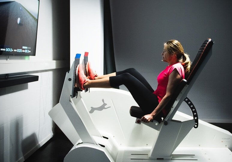
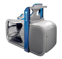
Finally, because problems in the hip often arise because of asymmetrical weight-bearing, we use Computer-Assisted Rehabilitation Environment, a breakthrough rehabilitation technology, to locate gait irregularities. C.A.R.E.N’s force-plate and motion-capture analysis creates a virtual-reality environment that measures motion of a subject and provides precise therapeutic strategies for recovery and healing.