Stroke Recovery and Treatment in New York
A stroke, much like a heart attack, is a condition involving obstruction of blood vessels. However, while a heart attack typically results from the prevention of blood circulation in the arteries, strokes are the prevention of blood circulation in the brain. Stroke is the third-leading cause of death in the United States and the second-leading cause of death worldwide. Those who survive may struggle with serious disability and impairment, which is why treatments like left-side stroke recovery and right-side stroke recovery are so crucial.
Vascular Brain Anatomy and Associated Conditions
Knowledge of the vascular anatomy, the network of blood vessels in the brain, can help us better determine the exact size and location of a stroke. This, in turn, is helpful in planning strategies for stroke patients’ recovery.
The brain is composed of the paired cerebral hemispheres (the cerebrum), the diencephalon, the brain stem, and the cerebellum.
The cerebrum is the largest portion of the brain, and contains a convoluted series of ridges. This region of the brain is divided into the bone that covers it.
The frontal lobe contains four gyri, or ridges. One gyrus (the pre-central gyrus) controls all the major movements of the body. The frontal eye fields control eye movements. The moto articulate in words what they’re capable of thinking.
The parietal lobe of the cerebrum is the primary sensory area, and is concerned with sensations like touch, temperature, and pain.
The occipital lobe primarily functions to the limbic system which helps manage emotions.
The diencephalon lies between the cerebrum and the brain stem, and is composed of the thalamus, hypothalamus, and epithalamus.
Like the cerebrum, the cerebellum is divided into the occipital and parietal lobes, the cerebellum coordinates balance and smooth motion of the musculoskeletal system.
Finally, the brain stem at the lower back of the brain consists of the midbrain, pons, and medulla oblongata. The superior colliculus of the midbrain is responsible for reflexes tory centers that regulate the heart and breathing.
The posterior portion of the brain receives blood from the basilar artery, a union of two vertebral arteries that pass through the cervical vertebrae and enter the cranium through a pass known as the foramen magnum. The basilar artery climbs along the brain stem and forms two posterior cerebral arteries. It’s these arteries that nourish the posterior portion of the cerebrum. Stroke affecting the basilar artery may result in dizziness, coma, headache, pinpoint pupils, quadriplegia (loss of movement in both arms and legs), and dysphagia (difficulty swallowing).
The superior cerebellar arteries branch from the basilar artery. Among the regions of the brain it supplies are the superior surface of the cerebellar hemispheres, most of the white matter in the cerebellum, and portions of the midbrain. The anterior inferior cerebellar artery branches from the basilar artery near the pons. Strokes that affect the superior cerebellar artery can cause ataxia, an inability to voluntarily coordinate muscle movements. Strokes that affect the anterior interior cerebellar artery can cause ataxia, sudden hearing loss, vertigo, facial paralysis, and Horner’s body syndrome (a condition of the sympathetic nervous system that results in drooping eyelid).
The anterior portion of the brain receives blood from the bilateral carotid arteries. The common carotid arteries pass through the neck along one side of the trachea. Just below the mandible, each of these arteries branches into the internal and external carotid arteries.
The internal carotid artery ascends the neck until it reaches the base of the neck and enters the brain. Once on the brain’s inferior surface, it splits into this artery from stroke can cause sensory loss, especially in the lower extremities, and mental confusion.
Importantly, the anterior and middle cerebral arteries provide blood to the cerebrum, and stroke affecting the middle cerebral artery can result in hemianopsia (loss of vision in half the visual field) and hemiparesis (weakness on one side of the body).
Strokes that affect the internal carotid artery result in hemianaesthesia (loss of sensation on one side of the face and on the opposite side of the body) and hemiplegia (partial or to that part of the carotid artery that lies in the cranium.
Types of Stroke and Post-Stroke Recovery
Left-brain stroke recovery focuses on the left half of the brain, which typically plays a more significant role in speaking ability than the right brain. (It should be noted that simplistic popular distinctions between the left brain and right brain are largely bogus). Aphasia is a common problem for people who have suffered a stroke on the left side of the brain, and patients with this condition may forget the right words in a given situation, speak in short, fragmented, or incomplete sentences, and have trouble comprehending conversations. They may also suffer from dyscalculia, a weakened ability to grasp mathematics, or apraxia, in which the performance of everyday tasks like putting on clothes is diminished. Some patients even struggle with amnesia and sudden changes in their emotional makeup.
Right-brain stroke recovery treats the side of the brain primarily associated with depth perception and visual-spatial functions. Patients who have suffered a stroke to register sarcasm. This, in turn, creates trouble in social situations.
Recovery from hemorrhagic stroke should begin immediately after stroke. A physical therapist may begin range-of-motion rehabilitative exercises in the hospital by moving a patient’s limb or having them move their own limb. Gradually a patient should returnto patient.
Stroke-Patient Recovery at the New York DNR
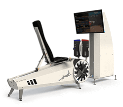
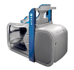
Patients seeking recovery from stroke will find a range of minor and major stroke recovery treatments at the New York DNR. We use advanced virtual reality and computer technology tore locomotion and balance.