Our Patient
Our patient is a 45 year-old female runner complaining of left sit bone pain when running and sitting. She has a history of proximal hamstring tendinopathy, and had been treated with plasma injections about 5 years prior, along with physical therapy. Her pain was initially resolved by the injections, and she continued to run.
The Challenge
The patient’s pain returned after 5 years, and she was treated with platelet rich plasma (PRP) injected into the proximal hamstring tendon under X Ray guidance. She initially experienced moderate improvement at six weeks after the injection, but her pain returned when she began to run again, along with sensations of tingling and tension spreading to the mid-thigh, and difficulty swinging her left leg.
Our Diagnostic Approach
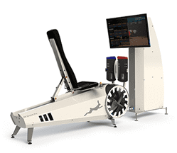
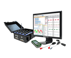
We conducted a clinical exam and a high-tech running gait analysis to detect the source of the patient’s pain and dysfunction. We also used diagnostic ultrasonography to visualize the patient’s hip and pelvic region in motion.
Our clinical exam revealed:
- Instability in the left sacroiliac joint
- Left hip instability
- Severely weakened muscles in the left gluteus medius, and loss of eccentric control of the deep intrinsic hip rotators
- Tenderness at and below the semimembranosus at its insertion to the sit bone
- A positive slump test (an assessment used to diagnose radicular pain originating from the lumbar spine)
- A positive test for ischio-femoral impingement
Our gait analysis revealed:
- A tendency to over-stride during running
- A shorter stride on the left leg
- Significant foot crossover of the left leg
- Left pelvic drop
Diagnostic ultrasonography revealed:
- Fraying of the left long dorsal sacroiliac ligament
- Thickening of the fascia in the thoracic-lumbar region, and fraying at its insertion to the left ilium
- Sciatic nerve splitting into tibial and peroneal branches at the piriformis muscle, with no evidence of nerve entrapment
- Significant fibrotic tissue lateral to the proximal hamstring insertion that was pushing the sciatic nerve towards the lesser trochanter.
- A significantly enlarged sciatic nerve with an abnormal shape, possibly representing an adhesion due to long-standing proximal tendinosis
- Abnormal movement of the hip, with the left lesser trochanter trending towards the sit bone
- Normal ischio-femoral space and normal-sized quadratus femoris
Our Diagnosis
Our diagnosis identified several issues:
- Severe proximal hamstring tendinopathy with scar tissue formation `
- Abnormal sciatic nerve mechanics
- Functional ischio-femoral impingement
- Sacroiliac joint dysfunction
Why the Patient’s Prior Treatments Failed
The patient’s initial treatment of platelet-rich plasma (PRP) injections five years prior successfully eradicated her symptoms, but no causative factors were established. The physical therapy she received was too general, and not specific to her problem. Since she kept running, her tendinopathy eventually returned and progressed to functional ischio-femoral impingement.
Her second PRP treatment also failed despite being done under Xray guidance, because soft tissues and nerves cannot be visualized with Xray. Her pain returned immediately when she began running again because the treatment addressed the symptoms without identifying the cause. The previous treatments were not ultrasound guided and did not address sciatic nerve mechanics.
In addition, the physical therapy she was receiving included exercises that were compressing the sciatic nerve and creating friction between her thickened hamstring tendon and the sciatic nerve. Such exercises combined with running forty miles a week eventually led to fibrosis of the sciatic nerve. Faulty ischio-femoral mechanics were overlooked, and gait retraining was not even considered.
Our Treatment Approach
- We performed ultrasound guided extracorporeal shockwave therapy (ESWT) on the proximal hamstring and sacro-illiac ligaments.
- We incorporated fascia manipulation to release fascia densifications along the retro-medial line.
- We used ultrasound guided dry needling in the deep sub-gluteal muscles, along the course of the sciatic nerve.
- Using TECAR therapy and manual mobilization, we improved the elasticity of the hip capsule.
- We used DNS therapy to promote hip stability.
- We retrained the patient’s running gait to eliminate over-striding.
- We provided a home exercise program for hip release and arch muscle training.
Our Results
The patient’s condition improved by 70% within 10 weeks. At that point, we referred her for sciatic nerve hydrodissection, and she was 100% symptom-free after two injections, and back to running to her normal mileage. She remained symptom-free at her 3-month checkup.
Conclusion
Treating symptoms without understanding their underlying cause can lead to further degradation of injured and/or dysfunctional tissues. A thorough and accurate diagnosis is key to rehabilitating patients and restoring pain-free mobility.