
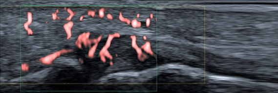
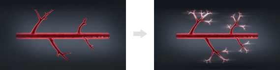
For any musculoskeletal injury or dysfunction, early and accurate diagnosis is key to speedy treatment and recovery. Superior microvascular imaging (SMI) is a highly sensitive and specific tool used in conjunction with grey-scale ultrasound imaging to diagnose pathological changes before they appear on any type of imaging. This is possible due to the ability of this breakthrough innovation to see otherwise invisible microscopic blood vessels.
This capability, in conjunction with grey scale high definition ultrasonography, provides the clinician with a 4th dimension view. It is not yet available with any other imaging modality or any other ultrasound technology. The importance of SMI for the rehab practitioner cannot be underestimated, as it not only allows for earlier and more precise diagnosis, but it has practical applications for treatment selection, monitoring of therapy dynamics, and pre- and post-treatment assessment.
Explore more advanced diagnostic tools available only at NYDNRehab:

Early and accurate diagnosis of injuries is key to speedy treatment and recovery. SMI (Superior microvascular imaging):

Verified Expert Profiles
Dr. Lev Kalika is a world-recognized expert in musculoskeletal medicine. with 20+ years of clinical experience in diagnostic musculoskeletal ultrasonography, rehabilitative sports medicine and conservative orthopedics. In addition to operating his clinical practice in Manhattan, he regularly publishes peer-reviewed research on ultrasound-guided therapies and procedures. He serves as a peer reviewer for Springer Nature.
Dr. Kalika is an esteemed member of multiple professional organizations, including:
Below is a prime example of how ultrasound can take the guesswork out of diagnosis.

A bad physical therapy experience is one of the primary causes of unnecessary surgery
In this instance, an athlete was originally diagnosed with minor quadriceps muscle strain and was treated for four weeks, with unsatisfactory results. When he came to our clinic, the muscle was not healing, and the patients’ muscle tissue had already begun to atrophy.
Upon examination using MSUS, we discovered that he had a full muscle thickness tear that had been overlooked by his previous provider. To mitigate damage and promote healing, surgery should have been performed immediately after the injury occurred. Because of misdiagnosis and inappropriate treatment, the patient now has permanent damage that cannot be corrected.
The most important advantage of Ultrasound over MRI imaging is its ability to zero in on the symptomatic region and obtain imaging, with active participation and feedback from the patient. Using dynamic MSUS, we can see what happens when patients contract their muscles, something that cannot be done with MRI. From a diagnostic perspective, this interaction is invaluable.
Dynamic ultrasonography examination demonstrating
the full thickness tear and already occurring muscle atrophy
due to misdiagnosis and not referring the patient
to proper diagnostic workup
Demonstration of how very small muscle defect is made and revealed
to be a complete tear with muscle contraction
under diagnostic sonography (not possible with MRI)

Complete tear of rectus femoris
with large hematoma (blood)

Separation of muscle ends due to tear elicited
on dynamic sonography examination


