Poor gait mechanics during running can dramatically reduce performance and cause overuse injuries. Over time, chronic pain sets in that is difficult to ignore. Many runners try to manage pain by wearing a knee brace, taping their ankles or taking NSAIDs. But chronic pain during and after running is not normal. Pain is your body’s way of alerting you that something is wrong.
Similarly, walking is a normal human activity that should be relatively effortless and pain free. Yet many people suffer pain and discomfort while walking. As humans age, walking often becomes difficult to the point of being dangerous. Loss of balance can lead to falls that result in serious fractures, painful contusions and head trauma.
Alterations in walking and running gait normally evolve and worsen over time. The changes are often so incremental that patients are not even aware of them until pain or an injury gets their attention. Gait analysis gathers quantitative data to help understand the underlying causes of gait abnormalities, That information can then be used to devise a treatment plan, with the goal of restoring healthy gait, eliminating pain and reducing injury risk.
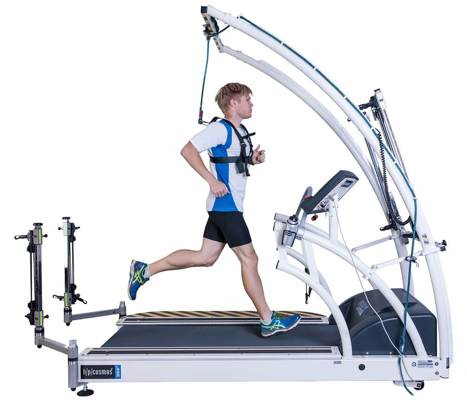
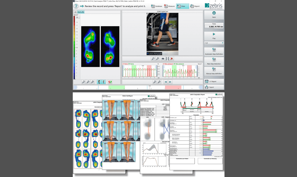
Today’s gait analysis is more precise and revealing than ever before, thanks to innovations in technology. We are now able to analyze gait in three dimensions to get a clearer picture of faulty gait patterns. Our process involves measuring various gait characteristics, identifying abnormal patterns and pinpointing the underlying cause.
At NYDNRehab, our gait analysis package includes a combination of technologies and methodologies unavailable elsewhere in New York:
- 3D gait analysis equipment and software to measure gait in three dimensions
- Real-time running feedback retraining
- Instrumented force treadmill to assess dynamic center of pressure
- Dual force plate technology to monitor bilateral ground reaction forces
- Diagnostic musculoskeletal ultrasonography
It is important to note that data collected with the most advanced technology is useless without an experienced team of technicians and clinicians to interpret and apply it. The sports medicine team at NYDNRehab has extensive experience with walking and running gait analysis and retraining. We know how to turn hard data into useful information that helps patients correct and restore healthy gait.
The clinic at NYDNRehab is one of few private clinics in the United States to employ sophisticated 3D gait analysis technology to diagnose and treat our patients. Our quantitative approach takes the guesswork out of gait analysis and gives us precise metrics to gauge the effectiveness of individualized treatment plans.
Anyone who walks or runs can benefit from 3D gait analysis and retraining. Pain and disability are not inevitable side effects of aging. The sooner you correct your gait mechanics, the better. Your quality of life, now and in the future, depends on it.
The data collected with 3D gait analysis is much more robust than 2D and provides a clinical certainty impossible to achieve with a 2D gait analysis system. Our clinic has the first 3D system to allow for real time running gait retraining.