Wrist Pain

Ongoing repetitive use of the wrist and hands can lead to painful conditions of the wrists that require wrist pain treatment .

Humans have always relied on strong wrists and hands to perform daily tasks like gardening, needlecraft, woodworking, building and just about any craft or trade you can think of. In modern times, technology has impacted the way in which most of us use our wrists and hands, and we often spend long hours typing on a computer keyboard or texting with our thumbs. In athletics, the wrists may be used to provide support, break a fall or stabilize the hand.

Anatomy of the Wrist

We commonly think of the wrist as a single joint, but it is actually a complex structure made up of multiple small joints that connect the carpal bones of the hand to the radius (on the thumb side) and the ulna (on the pinky side) of the forearm. While the structure of the wrist bears some similarity to the ankle, its bones are smaller and more fragile, with less cartilage and thinner ligaments.

The wrist is made up of eight carpal bones arranged in two rows of four. The ulna does not connect directly to the carpals, but its confluence is facilitated by a disc called the Triangular Fibrocartilage Complex (or TFCC) that serves as a sort of meniscus in the wrist. The TFCC can be easily injured during weight bearing activities. The TFCC also attaches to the radius though ligaments and fascia, so an injury can have a broad effect on wrist function.

wrist pain treatment nyc

Every small bone in the wrist forms a joint with adjacent small bones, creating dozens of joints that allow for a diverse range of movement and dexterity of the wrist and hand. Multiple ligaments throughout the wrist provide support to the wrist joints.

The muscles that provide wrist and hand movement are primarily located in the forearms, with long tendons that reach into the wrist and hand. Two thick ligamentous bands at the wrist anchor the tendons in place. The anterior, or palmar band, called the transverse carpal ligament, forms a tunnel with the carpals through which passes the median nerve that runs the length of the arm, into the hand.

icon

Why Physical Therapy Alone May Not Resolve Your Condition

Physical therapy is a valuable and effective approach to resolving musculoskeletal pain and dysfunction, but in many cases, physical therapy does not provide a stand-alone solution. Prior to beginning physical therapy, patients often need to address underlying issues that contribute to their pain and disability.

Unfortunately, mainstream physical therapy clinics are often not adequately equipped or experienced to identify and treat complications that undermine the effectiveness of physical therapy. They often rely on one-size-fits-all treatment protocols that overlook the unique characteristics of the individual condition, opting to treat the symptoms and not the patient.

Issues that should be addressed prior to beginning physical therapy include:

  • Scar tissue and fascia adhesions
  • Neurogenic inflammation
  • Joint edema
  • Inflamed soft tissues
  • Myofascial trigger points
  • Compressed or entrapped nerves
  • Tendons that have degenerated and lost their elastic properties
  • Compensation patterns developed post-injury
Block PT

Holistic Non-Invasive Diagnosis and Regenerative Therapy

Identifying and treating underlying issues prior to beginning physical therapy is key to getting fast and effective results. Failure to do so can completely undermine your treatment protocol, and in some cases, your condition may even worsen.

At NYDNRehab, we use a broad range of advanced technologies and innovative therapeutic approaches to resolve issues that can potentially undermine the success of physical therapy.

Our talented staff is certified in a diverse array of treatment methodologies, rarely found in run-of-the-mill physical therapy clinics. Our one-on-one sessions are personalized, based on the patient’s unique diagnostic profile.

Obstacles to physical therapy success include:

  • Scar tissue and fascia adhesions
  • Neurogenic inflammation
  • Joint edema
  • Inflamed soft tissues
  • Myofascial trigger points
  • Compressed or entrapped nerves
  • Tendons that have degenerated and lost their elastic properties
  • Compensation patterns developed post-injury

Common Conditions of the Wrist

Because of its complexity, there are many things that can cause wrist pain and reduced mobility. Some common conditions include:

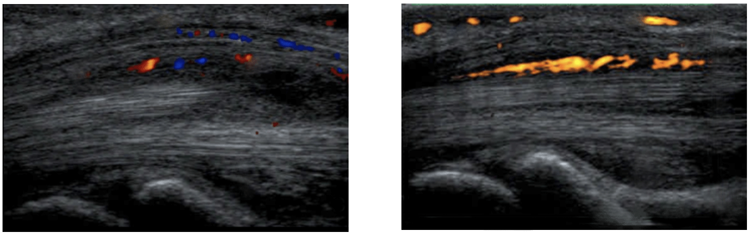
Carpal tunnel syndrome: This condition occurs when the median nerve becomes compressed within the carpal tunnel, often due to swelling or hypertrophy of the tendons that pass through the tunnel from the forearm. Pain may also arise from restricted gliding of the nerve anywhere along its pathway down the arm.

Ganglion cysts: A ganglion cyst is a fluid-filled lump that appears near the joints or tendons, often at the top or on the palm side of the wrist. Their cause is unknown, and they are sometimes but not always painful.

Osteoarthritis: Arthritis is caused by deterioration of the joint cartilage, marked by pain, stiffness and swelling. It has been linked to chronic systemic inflammation and metabolic disorders.

Thumb sprain: A sprain is injury to a ligament, most often the ulnar collateral ligament that connects the thumb to the hand. A thumb sprain is a common sports injury, and may also occur from a fall.

Trigger finger: Medically called stenosing tenosynovitis, trigger finger is often seen in patients with rheumatoid arthritis, gout and diabetes. It arises from a thickening of tissue that inhibits smooth gliding of tendons.

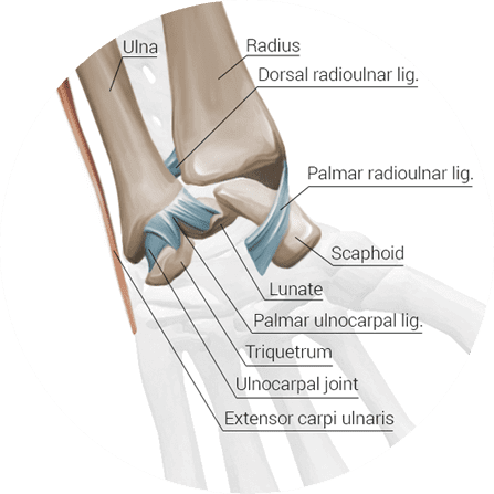
Wrist fractures: The wrist is made up of the eight carpals, along with the radius and ulna. Any of these bones can sustain a fracture, but the radius is the most common site of wrist fracture. Falls are the most common cause of wrist fractures. A fall may also do damage to ligaments, tendons, muscles and nerves.

Diagnosis of Wrist Pain

app

At NYDNRehab, we are experts in diagnostic musculoskeletal ultrasonography and neuro sonography. Our clinic features the highest resolution ultrasound machine in NYC, with a 24 MHz probe, SMI (superior microvascular imaging) and sonoelastography.

High resolution ultrasonography is far superior to MRI. It allows for dynamic imaging and the ability to quantify elasticity of the median nerve and other structures within the carpal tunnel. These features let us make a more precise diagnosis, choose the proper intervention and monitor the progress of treatment. Pre and post imaging can be performed multiple times during the course of treatment.

img

Please explore more advanced diagnostic option unavailable anywhere else:

img

Treatment of Wrist Pain

Treatment of your wrist pain will vary, depending on the source and nature of your condition. In most cases, conservative treatment with physical therapy and other non-invasive methods is sufficient to resolve pain and restore function.

Wrist Pain Diagnosis and Treatment at NYDNR

The wrist pain specialists at NYDNR take a multi-modal and holistic approach to treating your wrist pain. We use dynamic real-time diagnostic ultrasound to view your wrists structures and identify the source of pain. Treatment may include physical therapy, extracorporeal shock wave therapy (ESWT), manual nerve mobilization and neurodynamics therapy, acupuncture, ultrasound guided dry needling (UGDN), nerve mobilization, and other innovative treatment methods geared to restoring pain-free function.

wrist pain treatment

About the Author

Dr. Lev Kalika is clinical director of NYDNRehab, located in Manhattan. Lev Kalika is the author of multiple medical publications and research, and an international expert in the field of rehabilitative sonography, ultrasound guided dry needling and sports medicine Dr. Kalika works with athletes, runners, dancers and mainstream clients to relieve pain, rehabilitate injuries, enhance performance and minimize the risk of injuries. His clinic features some of the most technologically advanced equipment in the world, rarely found in a private clinic.

bg

In this instance, an athlete was originally diagnosed with minor quadriceps muscle strain and was treated for four weeks, with unsatisfactory results. When he came to our clinic, the muscle was not healing, and the patients’ muscle tissue had already begun to atrophy.

Upon examination using MSUS, we discovered that he had a full muscle thickness tear that had been overlooked by his previous provider. To mitigate damage and promote healing, surgery should have been performed immediately after the injury occurred. Because of misdiagnosis and inappropriate treatment, the patient now has permanent damage that cannot be corrected.

The most important advantage of Ultrasound over MRI imaging is its ability to zero in on the symptomatic region and obtain imaging, with active participation and feedback from the patient. Using dynamic MSUS, we can see what happens when patients contract their muscles, something that cannot be done with MRI. From a diagnostic perspective, this interaction is invaluable.

Dynamic ultrasonography examination demonstrating
the full thickness tear and already occurring muscle atrophy
due to misdiagnosis and not referring the patient
to proper diagnostic workup

Demonstration of how very small muscle defect is made and revealed
to be a complete tear with muscle contraction
under diagnostic sonography (not possible with MRI)

image

Complete tear of rectus femoris
with large hematoma (blood)

image

Separation of muscle ends due to tear elicited
on dynamic sonography examination

bg
Buy now 3D Gait
Payment Success
Request Telehealth Request Telehealth Request in office visit Book now