Most people take movement for granted until something goes wrong. Even then, many people accept reduced mobility as a natural part of aging. But the human body is designed to move in fluid and coordinated patterns that conserve energy, generate power, and optimize mechanical potential.
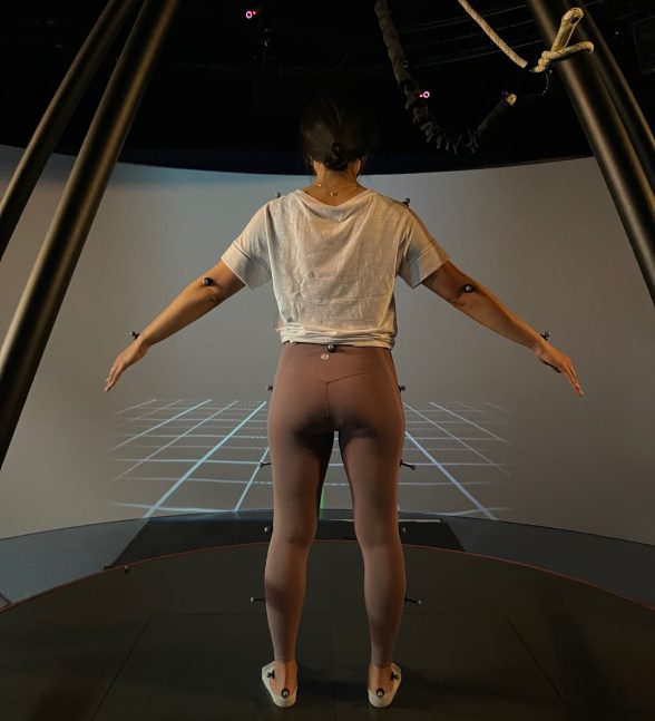
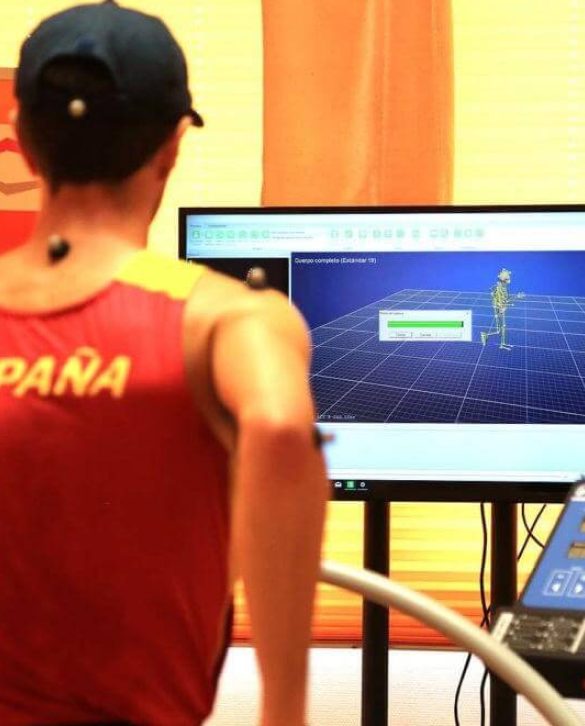
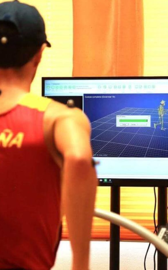
When movement is impaired by injury or overuse, a 3D motion analysis can help us accurately identify factors that impede movement and cause pain and dysfunction. Once we have a clear picture of the underlying causes of impaired movement, we are able to create a personalized treatment plan based on your unique patient profile.
USono technology allows us to visualize your body in motion via diagnostic ultrasound, and sync it with data from our cutting-edge motion analysis technology. Our 3D system is based on the latest evidence-based research on motion analysis.
or

Clinical director & DC RMSK
Verified Expert Profiles
NYDNRehab clinical director Dr. Lev Kalika has devoted his life’s work to revolutionizing the way physical pain syndromes and motor disorders are diagnosed and treated. Dr. Kalika is a world-renowned expert in diagnostic musculoskeletal ultrasonography, and has contributed multiple research articles to the growing body of scientific literature on its use in rehabilitative medicine.
The clinic at NYDNRehab features the highest resolution ultrasound equipment available for diagnosis and ultrasound-guided therapies. When paired with our motion capture system, ultrasound gives us crystal-clear images of your body in motion, in real time.
Our 3D motion analysis technology lets us accurately detect motor deficits that contribute to pain and reduced mobility. Prior to beginning physical therapy, we use our regenerative technologies to pre-treat damaged tissues, to stimulate and accelerate the healing process. Dr. Kalika is trained and certified in a broad range of rehabilitative treatment modalities. His ongoing commitment to personal growth and continuing education, along with his personalized approach to patient care, set NYDNRehab apart as the premier clinic for biomechanical analysis and rehabilitative therapy in NYC.The motion analysis lab at NYDNRehab features some of the most advanced technologies for biomechanical analysis currently available. Such technologies are rarely found in a private clinical setting.
Our motion analysis lab is ever-evolving as new research and technologies emerge. Our data-driven approach gives us measurable markers that ensure your treatment delivers the desired results. We designed our motion analysis lab based on SEBT research. SEBT is considered the gold standard for motion analysis protocols.
3D motion analysis is a full-body approach that registers motion in the lower and upper body. It provides data about movement in the following parameters:
3D Motion Analysis is appropriate for:
Humans are born with innate developmental software that governs their progression from helpless infant to active toddler. As human babies overcome gravity and master upright movement, they are able to run, jump and acquire new skills with ease.
Over time, certain factors may come into play that disrupt functional movement patterns, such as injuries, poor posture, environmental toxins, and other threats. 3D motion analysis can help people with impaired movement to identify and rehabilitate dysfunctional patterns and restore functional movement.
Much like the engine of your car, the human body is more than the sum of its parts — it is a complex organism made up of integrated and interdependent systems that all play a role in human movement. Isolating a single part of the body or zeroing in on symptoms alone to diagnose a patient’s condition inevitably leads to mistreatment or under treatment, often spinning a web of complexities that worsen the patient’s condition.
3D motion analysis lets us look beneath the hood, to view your body’s systems in motion and observe how they interact. It helps us determine whether your symptoms are causative or compensatory, and sheds light on complex problems that involve multiple structures.
A patient presents with prior ankle trauma that appears stable based on clinical testing and diagnostic ultrasonography. However, those diagnostic tools cannot really tell us the whole story. There may be underlying sensory-motor ankle instability that can alter movement along the entire kinetic chain, affecting the knee, hip, pelvis and lumbar spine.
In other words, prior trauma can create compensation patterns that are invisible to the naked eye. They can cause suboptimal and uncoordinated muscle firing patterns that affect other joints, creating a false diagnosis of ankle stability. 3D motion analysis helps us unravel the factors that cause pain and reduce mobility and stability.
For physically active people, getting a 3D motion analysis is akin to taking your car in for a tuneup — it lets you identify potential problems before they become major repairs. The motion analysis lab at NYDNRehab is second to none in NYC. Our state-of-the-art technologies and proprietary software put us head-and-shoulders above other Manhattan rehab clinics.
If you are ready to restore your body to the way it was designed to move, our motion analysis lab is the place to start. From there, we will customize a treatment plan based on your unique profile. Don’t resign yourself to a life of impaired mobility that keeps you from doing the things you love. Contact NYDRehab to schedule your 3D motion analysis today, and get ready to get moving again!

Below is a prime example of how ultrasound can take the guesswork out of diagnosis.

A bad physical therapy experience is one of the primary causes of unnecessary surgery
In this instance, an athlete was originally diagnosed with minor quadriceps muscle strain and was treated for four weeks, with unsatisfactory results. When he came to our clinic, the muscle was not healing, and the patients’ muscle tissue had already begun to atrophy.
Upon examination using MSUS, we discovered that he had a full muscle thickness tear that had been overlooked by his previous provider. To mitigate damage and promote healing, surgery should have been performed immediately after the injury occurred. Because of misdiagnosis and inappropriate treatment, the patient now has permanent damage that cannot be corrected.
The most important advantage of Ultrasound over MRI imaging is its ability to zero in on the symptomatic region and obtain imaging, with active participation and feedback from the patient. Using dynamic MSUS, we can see what happens when patients contract their muscles, something that cannot be done with MRI. From a diagnostic perspective, this interaction is invaluable.
Dynamic ultrasonography examination demonstrating
the full thickness tear and already occurring muscle atrophy
due to misdiagnosis and not referring the patient
to proper diagnostic workup
Demonstration of how very small muscle defect is made and revealed
to be a complete tear with muscle contraction
under diagnostic sonography (not possible with MRI)

Complete tear of rectus femoris
with large hematoma (blood)

Separation of muscle ends due to tear elicited
on dynamic sonography examination


