Anyone who has ever had an X-ray or MRI knows that the images from those procedures are still shots, taken while the patient is immobile. But the human body is a living moving organism, and pain often manifests during physical activity. What if there was a way to see inside the body to observe the muscles and joints in motion, while simultaneously getting feedback from the patient?
Real-time ultrasound imaging (RUSI) offers just such technology. It is a valuable diagnostic tool for visually observing musculoskeletal structures, including muscles, tendons, ligaments, joints and bursae as they interact in real-time, in ways the patient would normally use them.
or

Clinical director & DC RMSK
Verified Expert Profiles
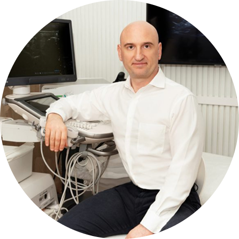
Dr. Lev Kalika, founder of NYDNRehab, has personally studied with some of the world’s most prestigious experts in diagnostic ultrasonography, including Carlo Martinolli MD,PhD , Alexander Kinzersky MD PhD , Thomas Clark DC, RMSK, Anna Vovchenko MD, and Rostislav Bubnov MD, PhD. Over the past seven years, Dr. Kaika has attended MUSOC, EFSUMB, AIUM and Gulfcoast Ultrasound Institute. He is an active member of the American Institute of Ultrasound in Medicine (AIUM), and is currently developing his own unique approach tors.
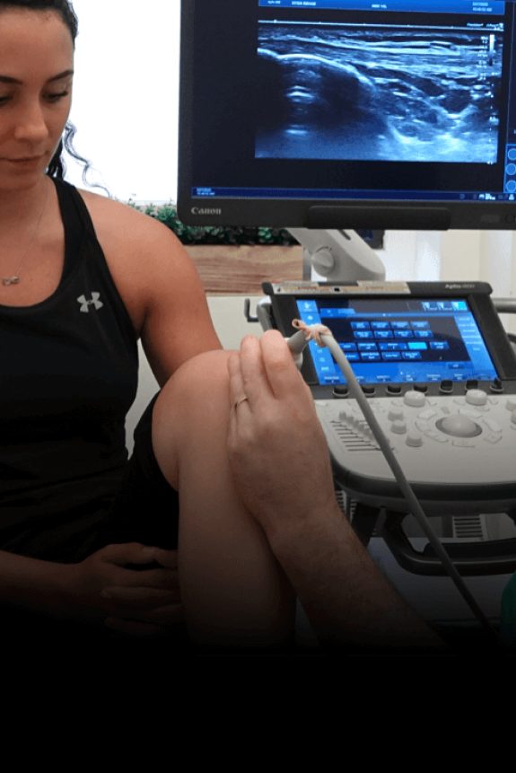
Dr. Kalika’s services are frequently sought out by elite athletes for rehabilitation and performance enhancement.During a RUSI session, a water-based gel is applied to the area being examined. A small transducer, or probe, is placed directly on the skin and transmits high-frequency sound waves through the gel into the body’s tissues. Sound waves are then reflected off the tissues. Because different tissues have different densities, the sound waves reflect back at various rates, making it possible to distinguish one type of tissue from another.
The transducer collects the sounds that bounce back, and a computer then creates images that are displayed on a screen. Because ultrasound images are captured in real-time, they can show the structure and movement of the body’s muscles, joints, internal organs, and even blood flowing through blood vessels.
Using RUSI, your therapist is able to observe the movements of your muscles as they occur and determine whether they are functioning correctly. The therapist can observe different layers of muscles contracting and relaxing, can look at the timing and size of muscle contractions, and can even see fatty tissue within your muscles.
RUSI is used in conjunction with an integrated total body assessment to diagnose muscle dysfunction. Feedback from RUSI can then be used to retrain muscles to perform optimal contractions. Images of a normal functional contraction can be compared to a patient’s dysfunctional muscle contraction so they are better able to understand the deficiency and how to correct it.

Real-time ultrasound imaging (RUSI) started to appear in the early 1980s, and by the1990s, 3D and 4D images were clear enough for the public to interpret, and were enhanced by color. Early iterations of the technology required large scan heads and heavy cables, making the equipment cumbersome and immobile. Today, some ultrasound equipment is so compact that it can be carried to patients on a battlefield or used by astronauts in space.
Since the 1980s, the use of RUSI for diagnosis and treatment of musculoskeletal injuries has continued to grow, as real-time ultrasound has numerous advantages over other imaging technologies like X-ray, MRI and CT scan:
As a case in point, isokinetic dynamometry is a tool commonly used to test peak muscle torque in research and rehabilitation. However, the method yields a relatively crude estimation of forces, and is unable to isolate individual muscles within a muscle group. For example, the quadriceps is a group of four distinct muscles that work together to produce movement. Isokinetic testing is not able to distinguish between the individual muscles, and only tests forces produced by the group as a whole. Yet when diagnosing and treating knee injuries such as ACL and meniscus tears, patelafemoral dysfunction, jumper’s knee and other injuries, it is important to isolate and test the function of individual muscles in the quadriceps. Real-time ultrasound imaging enables us to view and measure the strength and function of each muscle with the knee in multiple positions, something that is not possible with isokinetic testing.


The vast applications of RUSI are growing daily, both for diagnosis and treatment. Patients with acute injuries, overuse syndromes, gait and performance deficiencies, and chronic diseases can all benefit from RUSI.
Physical therapists can use RUSI to provide quick diagnosis and perform muscle re-education through visual feedback in a variety of patients, including:Virtually anyone who suffers from muscle and joint pain, dysfunction or instability can benefit from RUSI.
It would be nice if athletic injuries were simple, clean and easy to multiple structures. Traditionally, X-Ray and MRI have been used to be read and interpreted. Meanwhile, athletes and coaches are left guessing about the extent and severity of an injury, and treatment is delayed.
When it comes to view with the patient in a static supine posture.
During movement, the deep core muscles play a crucial role in mediating load transfer between the lower and upper body. When the core muscles are weakened, the pelvis becomes less stable, increasing the risk of injuries, low back pain and herniated discs. Real time ultrasonography has been shown in multiple clinical trials to collaborate in the diagnostic process, with the body in motion.
Musculoskeletal Ultrasound (MSUS) delivers high frequency sound waves to be done within minutes after the injury occurs. Moreover, the images can be viewed in real time by both clinician and patient.
Advantages of MSUS over X-Ray and MRI include:
For athletes, early and accurate diagnosis and treatment means sooner return to play. Some common athletic injuries and pathologies for which MSUS provides superior imaging include:


Research authored or co-authored by the clinic’s medical director. The following research publications inform the clinical approach used in this treatment program.
Conference Abstract
2024
 Lev Kalika
  Rostyslav Bubnov
Below is a prime example of how ultrasound can take the guesswork out of diagnosis.

A bad physical therapy experience is one of the primary causes of unnecessary surgery
In this instance, an athlete was originally diagnosed with minor quadriceps muscle strain and was treated for four weeks, with unsatisfactory results. When he came to our clinic, the muscle was not healing, and the patients’ muscle tissue had already begun to atrophy.
Upon examination using MSUS, we discovered that he had a full muscle thickness tear that had been overlooked by his previous provider. To mitigate damage and promote healing, surgery should have been performed immediately after the injury occurred. Because of misdiagnosis and inappropriate treatment, the patient now has permanent damage that cannot be corrected.
The most important advantage of Ultrasound over MRI imaging is its ability to zero in on the symptomatic region and obtain imaging, with active participation and feedback from the patient. Using dynamic MSUS, we can see what happens when patients contract their muscles, something that cannot be done with MRI. From a diagnostic perspective, this interaction is invaluable.
Dynamic ultrasonography examination demonstrating
the full thickness tear and already occurring muscle atrophy
due to misdiagnosis and not referring the patient
to proper diagnostic workup
Demonstration of how very small muscle defect is made and revealed
to be a complete tear with muscle contraction
under diagnostic sonography (not possible with MRI)

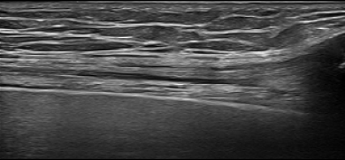
Complete tear of rectus femoris
with large hematoma (blood)

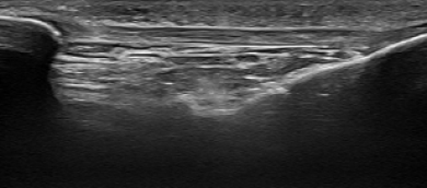
Separation of muscle ends due to tear elicited
on dynamic sonography examination