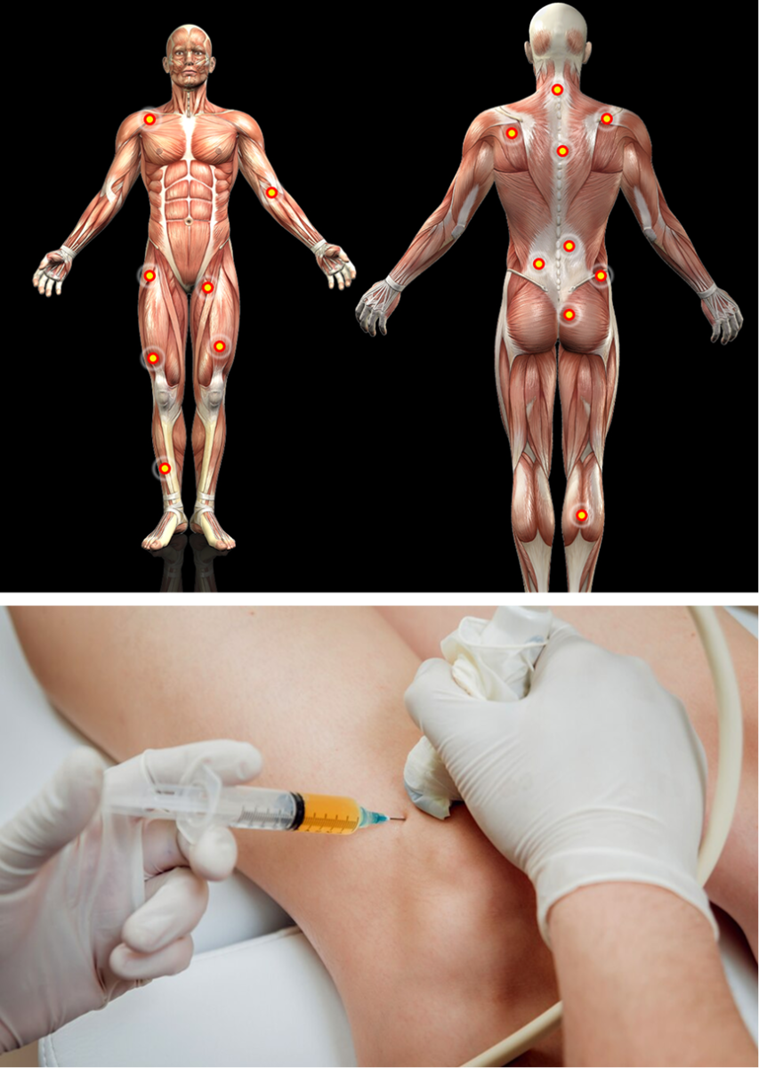
Soft tissue injuries are common in athletes and physically active people. They arise over time from microtrauma to soft tissues when they are forcefully loaded again and again, without adequate time for recovery. Injuries also occur in sedentary populations whose structures are too weak to support and stabilize the joints. If left untreated, soft tissue injuries can lead to conditions like tendinopathy, joint instability, and osteoarthritis, resulting in a diminishing quality of life and loss of independence.
When overuse injuries cause chronic tendon damage (tendinopathy) or damage to the boney insertion sites of tendons and ligaments (enthesopathy) they can begin to degenerate, leading to reduced performance, chronic pain, and physical dysfunction. When fascia is injured, it can densify and lose its elastic and gliding properties, causing loss of tensegrity and changes in muscle firing patterns.
Prolotherapy is an evidence-based therapeutic treatment approach that stimulates the healing of chronic soft tissue injuries and degenerative joint disorders. When appropriately applied, Prolotherapy can help to restore functional mobility, enhance performance and improve quality of life.

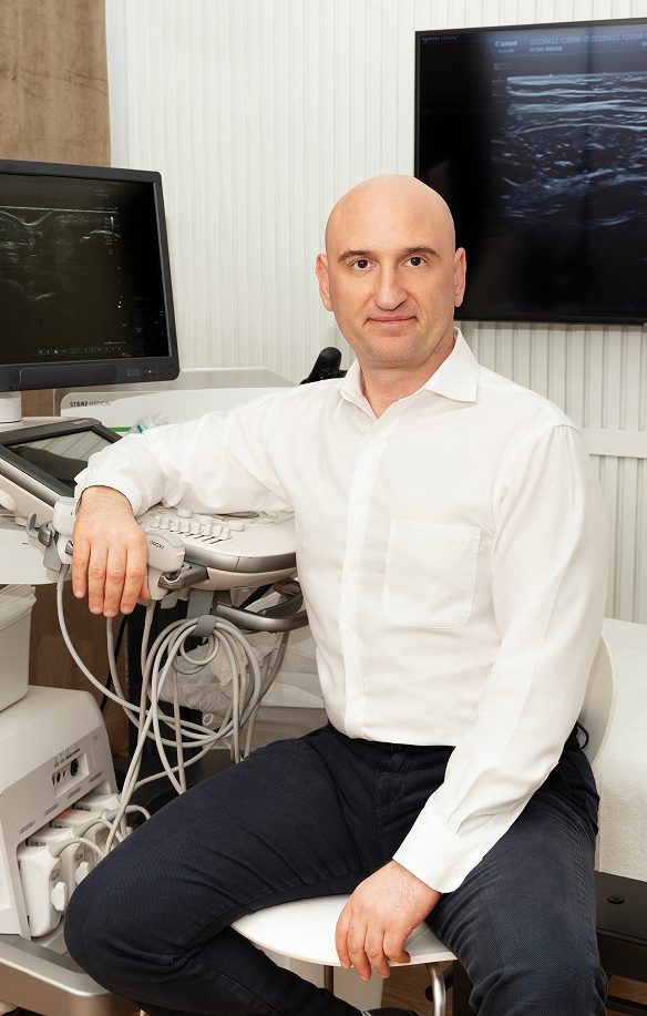
Orthobiologic, Sports Medicine and Regenerative Medicine Specialist
Since the inception of Prolotherapy some 30 years ago, we have seen incredible scientific advancements in the fields of soft tissue science and ultrasonography. Yet despite the emergence of new technologies and volumes of new research, most doctors are still using the outdated Hackett-Hemwall approach that involves “blind” needling into the locus of pain, without ultrasound guidance.
The conventional approach assumes that the patient’s body conforms to the standard human anatomical model, but we know that the architecture of anatomical structures can vary greatly from one patient to the next. Without dynamic high-resolution imaging, it is impossible to precisely target specific tissues.
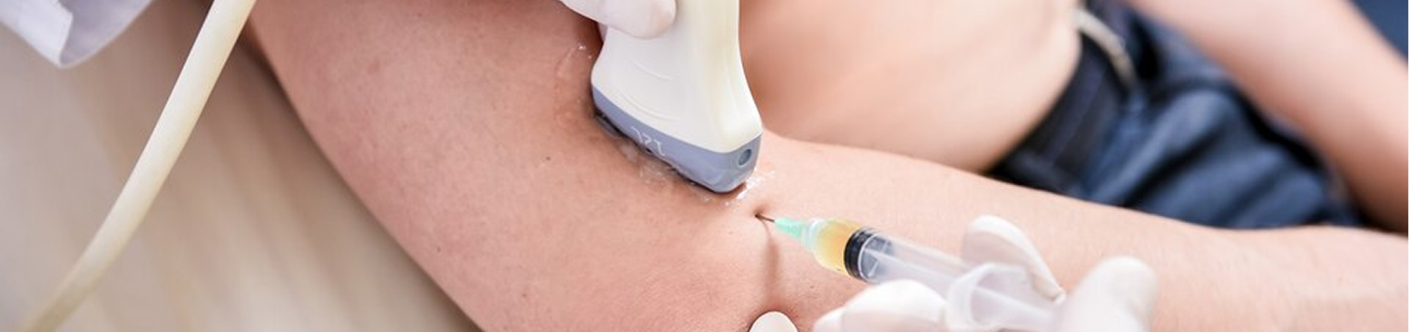
Prolotherapy involves injecting a neutral solution – most often dextrose-based – into injured or weakened ligaments, tendons, fascia, and joints, causing a mild inflammatory response in the targeted tissues. The inflammation attracts growth factors to the site that stimulate the body’s natural healing processes, triggering the production of new collagen to repair and strengthen injured tissues.
Most injuries involve multiple tissue types, and different tissues respond differently to Prolotherapy injections. It is not enough to blindly inject a solution at the locus of pain and hope that it has a regenerative effect. Without real-time ultrasound guidance, the procedure is hit-or-miss, which can dramatically limit its effectiveness.
At NYDNRehab, we use the highest-resolution ultrasonography to identify and differentiate various tissue types. We customize the concentration and volume of the Prolotherapy solution, based on the tissues involved. We then conduct our needling procedure under ultrasound guidance, to precisely target specific tissues for the most effective results. Our approach using advanced technologies is redefining how Prolotherapy are used and administered.
“The soul of man, with all the streams of pure living water, seems to dwell in the fascia of his body” – Andrew Taylor Still, MD, founder of osteopathy
Prolotherapy treatment is designed to strengthen different types of connective tissues like tendons, ligaments and fascia, to improve mobility and stability, but it would be irresponsible to administer needling therapies like Prolotherapy, PRP injections without first optimizing the tissues for treatment.
To function optimally, the locomotor system relies heavily on stability generated by myofascial tensegrity. But damaged fascia can form densifications and adhesions that pull against newly reinforced tissues, causing them to fray or rupture. Prior to Prolotherapy treatment, fascial distortions must be addressed.
At NYDNRehab we pretreat tissues with the following therapies:
If tissues are not prepared prior to Prolotherapy, the intended results may be difficult to achieve, requiring additional and more painful interventions.
Accurate and effective needling procedures require advanced knowledge of human anatomy, derived from cadaver dissection studies and the use of high-resolution sonography. But knowledge of human anatomy is not enough. According to recent research, and based on our own experience, prolotherapy injections performed blindly can do more harm than good.
To be effective, Injection therapies need to precisely target specific tissues with the appropriate volume of solution. Without ultrasound guidance, it is impossible to know how much of the injected solution is required, or if it has successfully hit its mark. And even that may not be enough, because ultrasonography itself requires extensive study and experience, and guided injection procedures require the highest-resolution ultrasound equipment available.
When a prolotherapy solution is injected blindly into the intermuscular plane, it is impossible to know how much solution is enough. Practitioners typically rely on outdated standardized guidelines defined by the Hamlet and Hackett protocol, which is not based on precise imaging and testing, and which often recommends a much greater volume of solution than is needed. We may see temporary pain relief and improved tissue gliding, but instead of enhancing tensegrity, we end up with fibrosis, fascial densifications, and adhesions that cause long-term issues.
At NYDNRehab, our personalized holistic approach is redefining how regenerative and orthobiologic therapies are used. Prior to prolotherapy injections, we conduct rigorous clinical testing to determine which tissues will benefit, and whether there is any related loss of tensegrity or fascial gliding.
In cases where Prolotherapy is not immediately warranted, we may opt to start with physical therapy, fascial manipulation, or other regenerative therapies. By contrast, many pain management doctors simply inject the solution into the locus of pain, without considering that the pain may arise from other causes that require different types of treatment.
In instances where fascia is densified and adheres to other structures, ultrasound enables us to combine classic Prolotherapy with interfascial plane injections – a procedure where a saline or dextrose solution is injected into the extracellular matrix between organized fascia layers, to separate soft tissues and restore functional movement.
We integrate all of our regenerative injection procedures with fascial manipulation and ultrasound-guided fascial hydro release. When combined with the most advanced high-resolution ultrasound imaging, we are able to visualize minute details, to determine the appropriate solution concentration, and ensure that the injections reach their intended target.
“Fascia is the fabric of life, the structure and flow between all cells (the extracellular matrix) and that which sustains the processes upholding connection, communication and interaction between all parts of the body. It constitutes the basic structural architecture of the human body.” – Fascia and the Living Body
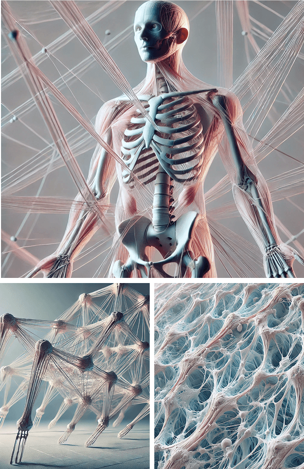
The integrity of your body’s structures and their capacity for optimal alignment and interaction is dependent on tension generated from your muscles and fascia – a phenomenon called biotensegrity. Your skeleton provides a structural framework for your entire organism, but without tension produced by muscles and fascia pulling against rigid bones, you would collapse in a motionless heap.
Myofascial tension must be tuned and balanced by strong and elastic tissues that allow the body’s structures to glide freely. Physical activity is essential for achieving and sustaining balanced myofascial tension, and athletes and physically active people rely on biotensegrity for peak performance. When the capacity of muscles and fascia to stretch, contract and glide is impaired, performance suffers and injury risk increases.
Loss of tensegrity, aka dystensegrity, is mostly mechanical in nature, arising from load transfer and distribution among muscles, ligaments, bones and viscera during physical activity. However, poor nutrition, inadequate hydration, prescription drugs and environmental toxins can also contribute to the deterioration of soft tissues.
Biotensegrity cannot be accurately assessed via a clinical exam or static imaging. Only dynamic high-resolution ultrasonography can give us real-time imaging of the myofascia in motion. Ultrasound lets us detect and measure fascia densifications and adhesions that contribute to dystensegrity. Sonoelastography gives us capabilities for measuring tension within myofascial tissues.
Our holistic and personalized approach to patient care addresses the many factors that undermine biotensegrity. Our regenerative technologies provide us with the tools we need to optimize performance and restore pain-free functional movement.
Chronic soft tissue injuries are often misdiagnosed, leading to inferior treatment approaches that waste patients’ time and money, while the tissues continue to deteriorate. At NYDNRehab, we know that the rehabilitation of chronic injuries takes time, and we don’t believe in wasting time on ineffective treatment.
We use the highest resolution diagnostic ultrasonography to dynamically visualize your injured tissues in real time. Ultrasound lets us differentiate between different tissue types and identify the nature, scope and severity of damage. Sonoelastography equips us to assess the density and elasticity of myofascial tissues, and superb microvascular imaging lets us monitor the healing process.
Chronic soft tissue injuries can be slow to heal, taking you away from your favorite activities for months on end. For athletes, chronic injuries can be career-ending and life-altering. For older adults, dystensegrity can cause conditions like joint osteoarthritis and chronic instability that severely reduces their quality of life.
Prolotherapy are important tools for rehabilitating chronic injuries and myofascial dysfunction. When combined with other advanced therapies and regenerative technologies, they provide our patients with the best treatment options available. Our commitment to excellence ensures that you receive the very best care, and recover in the least amount of time possible. Contact NYDNRehab today, and let us put you on the road to injury recovery so you can get back out there!


Independent peer-reviewed research relevant to this treatment approach.
Research authored or co-authored by the clinic’s medical director. The following research publications inform the clinical approach used in this treatment program.
Conference Abstract
2022
 Lev Kalika
  Rostyslav Bubnov
 Conference Abstract
2023
Lev Kalika Rostyslav Bubnov Conference Abstract
2023
Lev Kalika Rostyslav Bubnov Conference Abstract
2022
Lev Kalika Rostyslav Bubnov Conference Abstract
2024
Lev Kalika Rostyslav Bubnov Conference Abstract
2025
Lev Kalika Rostyslav Bubnov 
Below is a prime example of how ultrasound can take the guesswork out of diagnosis.

A bad physical therapy experience is one of the primary causes of unnecessary surgery
In this instance, an athlete was originally diagnosed with minor quadriceps muscle strain and was treated for four weeks, with unsatisfactory results. When he came to our clinic, the muscle was not healing, and the patients’ muscle tissue had already begun to atrophy.
Upon examination using MSUS, we discovered that he had a full muscle thickness tear that had been overlooked by his previous provider. To mitigate damage and promote healing, surgery should have been performed immediately after the injury occurred. Because of misdiagnosis and inappropriate treatment, the patient now has permanent damage that cannot be corrected.
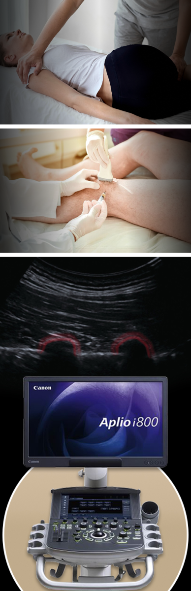
The most important advantage of Ultrasound over MRI imaging is its ability to zero in on the symptomatic region and obtain imaging, with active participation and feedback from the patient. Using dynamic MSUS, we can see what happens when patients contract their muscles, something that cannot be done with MRI. From a diagnostic perspective, this interaction is invaluable.
Dynamic ultrasonography examination demonstrating
the full thickness tear and already occurring muscle atrophy
due to misdiagnosis and not referring the patient
to proper diagnostic workup
Demonstration of how very small muscle defect is made and revealed
to be a complete tear with muscle contraction
under diagnostic sonography (not possible with MRI)

Complete tear of rectus femoris
with large hematoma (blood)

Separation of muscle ends due to tear elicited
on dynamic sonography examination


