
Muscle and soft tissue injuries are common among athletes from novice to professional, and the myofascial system plays an important role in the healing process. The myofascia provides support for muscle fibers and transmits mechanical signals between muscles and tendons. Real-time sonoelastography give us accurate information about the state of soft tissues, the progression of muscle healing, local muscle elasticity, and the stages of the myofascia repair process.

The myofascia, sometimes referred to as the “skeleton” of muscle fibers, forms a 3D network of dense tissue that surrounds and connects the musculoskeletal system.
Myofascia consists of three distinct layers of connective tissue:
Injuries occur not just to muscle fibers, but also to myofascia at the endomysium, perimysium and epimysium levels. When myofascia is injured, myogenic satellite cells fuse with healthy muscle cells to repair injured tissues and stimulate the growth of muscle fibers.
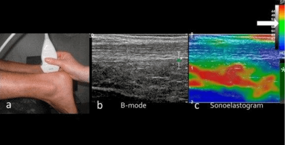
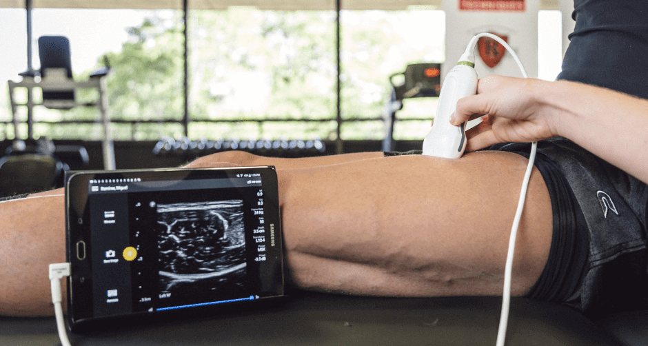
Soft tissues like muscles, subcutaneous fat and connective tissues have varying amounts of elasticity, or deformability, which is determined by the tissues’ composition and structure. Ultrasound imaging provides a safe, inexpensive and convenient way to view soft tissues in real time, with the patient in motion. Sonoelastography (SEL) works in conjunction with ultrasound imaging to assess the mechanical and elastic properties of soft tissues.
Soft tissue elasticity is expressed as a ratio of stress to strain. Injured tissues are typically harder and less elastic than normal healthy tissues. Soft tissue elasticity can be altered by tendinopathies, neuromuscular disorders and during the various stages of wound healing. SEL allows practitioners to assess muscle injuries based on elasticity, and gauge the progression of healing throughout the course of treatment.
Sonoelastography (SEL) can help us detect subtle changes in the mechanical properties of muscles, tendons, fascia and nerves in the early phases of injury. Early detection helps practitioners devise the most effective rehab strategies. SEL helps us monitor treatment progress and healing, enhance rehab protocols, eliminate pain, restore function, speed recovery and improve performance. With SEL, we can quantify muscle, tendon and nerve stiffness, giving us important data about the state of an injury and its healing progress.
Being able to quantify tissue properties has important repercussions for return to sport and injury prevention. Returning to the playing field prematurely after an injury dramatically increases an athlete’s risk of re-injury. Rather than basing rehabilitation progression and return to play on an arbitrary timeline, we are able to assess each injury and each athlete on a case by case basis.
There is no question that technology is making a profound impact on athletic performance. The ability to gather quantitative data on an individual athlete and apply it to training and rehab helps us build stronger athletes with more precise skills execution and lower risk of injury.
At NYPDNRehab, we have the latest and most sophisticated technologies at our disposal. Our unique technological toolkit includes:
Whether you have acute or repetitive overuse injuries, are recovering from surgery, suffer from chronic muscle or joint pain, or simply want to gain a competitive edge, the sports medicine team at NYDNRehab will help you optimize your musculoskeletal system and reach your peak performance potential.


Verified Expert Profiles
Dr. Lev Kalika is a world-recognized expert in musculoskeletal medicine. with 20+ years of clinical experience in diagnostic musculoskeletal ultrasonography, rehabilitative sports medicine and conservative orthopedics. In addition to operating his clinical practice in Manhattan, he regularly publishes peer-reviewed research on ultrasound-guided therapies and procedures. He serves as a peer reviewer for Springer Nature.
Dr. Kalika is an esteemed member of multiple professional organizations, including:
Below is a prime example of how ultrasound can take the guesswork out of diagnosis.

A bad physical therapy experience is one of the primary causes of unnecessary surgery
In this instance, an athlete was originally diagnosed with minor quadriceps muscle strain and was treated for four weeks, with unsatisfactory results. When he came to our clinic, the muscle was not healing, and the patients’ muscle tissue had already begun to atrophy.
Upon examination using MSUS, we discovered that he had a full muscle thickness tear that had been overlooked by his previous provider. To mitigate damage and promote healing, surgery should have been performed immediately after the injury occurred. Because of misdiagnosis and inappropriate treatment, the patient now has permanent damage that cannot be corrected.
The most important advantage of Ultrasound over MRI imaging is its ability to zero in on the symptomatic region and obtain imaging, with active participation and feedback from the patient. Using dynamic MSUS, we can see what happens when patients contract their muscles, something that cannot be done with MRI. From a diagnostic perspective, this interaction is invaluable.
Dynamic ultrasonography examination demonstrating
the full thickness tear and already occurring muscle atrophy
due to misdiagnosis and not referring the patient
to proper diagnostic workup
Demonstration of how very small muscle defect is made and revealed
to be a complete tear with muscle contraction
under diagnostic sonography (not possible with MRI)

Complete tear of rectus femoris
with large hematoma (blood)

Separation of muscle ends due to tear elicited
on dynamic sonography examination


