Case Study: Ultrasound Reveals Sciatic Neuropathy and Life-Threatening Peripheral Vascular Disease
Our Patient
Our patient is a 62-year-old female complaining of leg pain, calf and foot muscle weakness, and sensations of numbness in her lower extremities.
Our Diagnostic Approach
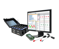
Our initial clinical exam indicated neuropathy, which we confirmed with multiple EMG tests. We followed up with an examination by high-resolution diagnostic ultrasonography.
Our ultrasound exam of the sciatic nerves revealed the following:
The size of the tibial and peroneal portions was consistent on the right and left sides.
Signs of sciatic neuropathy were more prominent on the right side.
Ultrasound imaging also revealed alarming muscle and vascular anomalies:
Muscle atrophy (shrinkage) in the right calf and decreased pennation angle of the muscle fibers (15 degrees on the right, compared to 23 degrees on the left).
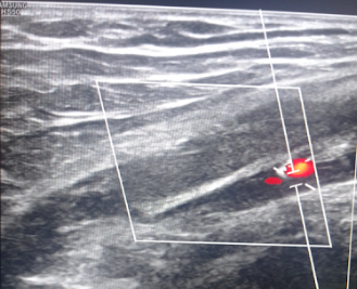
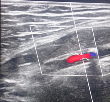
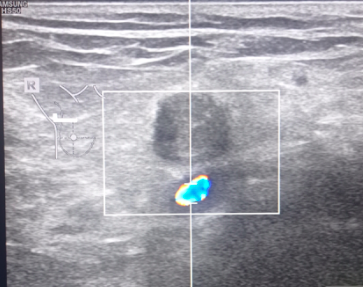
Complete occlusion (blockage) of the right popliteal artery.
Complete occlusion of the right femoral artery at the level of the inguinal ligament.
Narrowing of the right posterior tibial artery due to arterial wall deposits.
Near-total occlusion of the left popliteal artery.
Evidence of sporadic calcified plaques in the affected arteries.
Our Solution
We recommended that our patient consult with a vascular surgeon, as her condition was dire and beyond the scope of our practice.
Conclusion
Had we based our diagnosis on the patient’s symptoms and our clinical exam alone, we would not have been alerted to her peripheral vascular disease. In this case, our high-resolution diagnostic ultrasonography was able to detect critical vascular anomalies that might have been overlooked until the patient suffered a life-threatening cardiovascular event.
In this instance, an athlete was originally diagnosed with minor quadriceps muscle strain and was treated for four weeks, with unsatisfactory results. When he came to our clinic, the muscle was not healing, and the patients’ muscle tissue had already begun to atrophy.
Upon examination using MSUS, we discovered that he had a full muscle thickness tear that had been overlooked by his previous provider. To mitigate damage and promote healing, surgery should have been performed immediately after the injury occurred. Because of misdiagnosis and inappropriate treatment, the patient now has permanent damage that cannot be corrected.
The most important advantage of Ultrasound over MRI imaging is its ability to zero in on the symptomatic region and obtain imaging, with active participation and feedback from the patient. Using dynamic MSUS, we can see what happens when patients contract their muscles, something that cannot be done with MRI. From a diagnostic perspective, this interaction is invaluable.
Dynamic ultrasonography examination demonstrating the full thickness tear and already occurring muscle atrophy due to misdiagnosis and not referring the patient to proper diagnostic workup
Demonstration of how very small muscle defect is made and revealed to be a complete tear with muscle contraction under diagnostic sonography (not possible with MRI)
Complete tear of rectus femoris with large hematoma (blood)
Separation of muscle ends due to tear elicited on dynamic sonography examination