Our patient is a middle-aged female with shoulder pain lasting 8 months. There was no history of trauma, and she had not been able to get a concrete diagnosis or treatment.
The patient had been seen at a reputable clinic and by different practitioners. She brought with her MRI and ultrasound reports with nonspecific findings, and none of her records cited a concrete diagnosis.
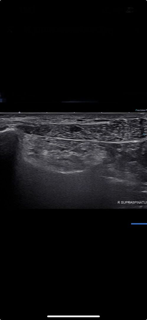
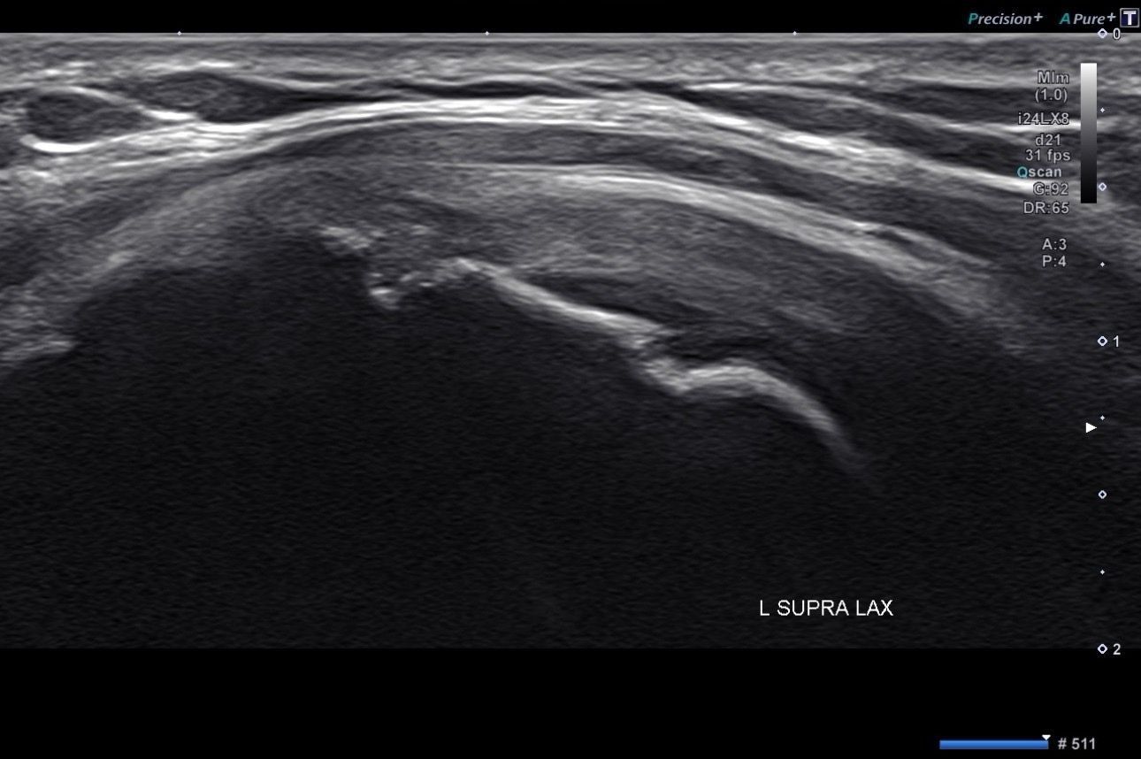
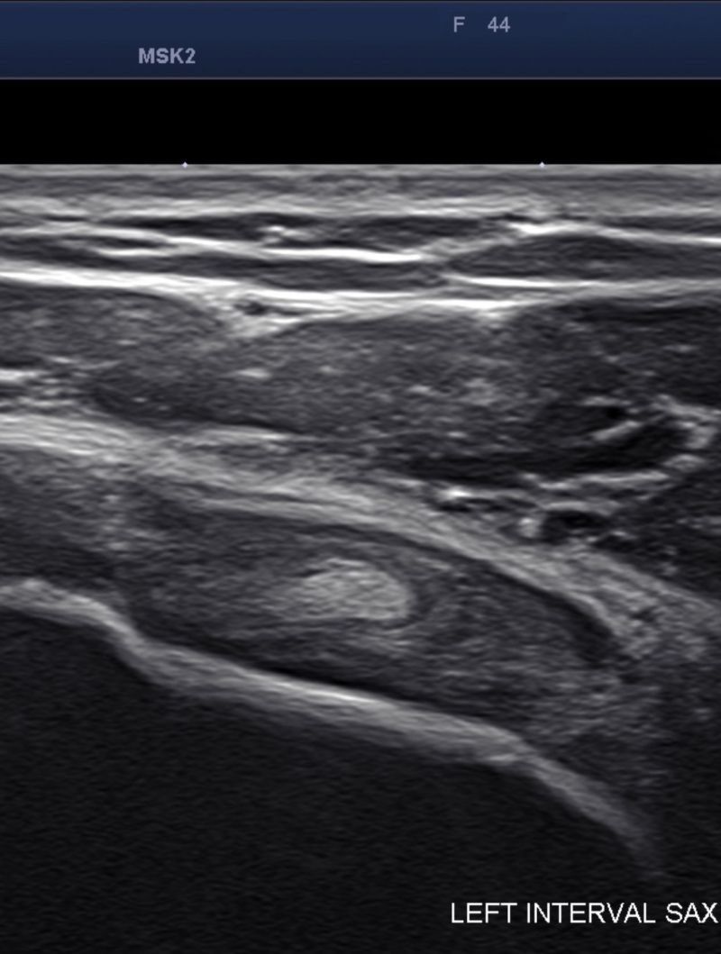
Our physical exam demonstrated severely restricted range of motion during abduction, internal and external rotation, indicating a clear case of adhesive capsulitis. We dove deeper to look for a thickened capsule in the axillary recess, examined the coracohumeral ligament, and looked for a biceps effusion – excess fluid within the long biceps tendon sheath – but found only mild hyperemia in the rotator interval
The patient was successfully treated using the following protocol:
The patient’s pain was alleviated with just one injection, and her shoulder range of motion was completely restored after just a few sessions.
The absence of findings from imaging does not rule out a clinical diagnosis, and some practitioners are over-reliant on imaging results. Also many practitioners see adhesive capsulitis as a self-limiting condition that resolves itself over time, and don’t feel treatment is necessary.
The patient suffered needlessly for 8 months when a single injection was enough to eliminate her pain, and her range of motion was restored with just a few regenerative energy sessions.

Verified Expert Profiles
Dr. Lev Kalika is a world-recognized expert in musculoskeletal medicine. with 20+ years of clinical experience in diagnostic musculoskeletal ultrasonography, rehabilitative sports medicine and conservative orthopedics. In addition to operating his clinical practice in Manhattan, he regularly publishes peer-reviewed research on ultrasound-guided therapies and procedures. He serves as a peer reviewer for Springer Nature.
Dr. Kalika is an esteemed member of multiple professional organizations, including:
Below is a prime example of how ultrasound can take the guesswork out of diagnosis.

A bad physical therapy experience is one of the primary causes of unnecessary surgery
In this instance, an athlete was originally diagnosed with minor quadriceps muscle strain and was treated for four weeks, with unsatisfactory results. When he came to our clinic, the muscle was not healing, and the patients’ muscle tissue had already begun to atrophy.
Upon examination using MSUS, we discovered that he had a full muscle thickness tear that had been overlooked by his previous provider. To mitigate damage and promote healing, surgery should have been performed immediately after the injury occurred. Because of misdiagnosis and inappropriate treatment, the patient now has permanent damage that cannot be corrected.
The most important advantage of Ultrasound over MRI imaging is its ability to zero in on the symptomatic region and obtain imaging, with active participation and feedback from the patient. Using dynamic MSUS, we can see what happens when patients contract their muscles, something that cannot be done with MRI. From a diagnostic perspective, this interaction is invaluable.
Dynamic ultrasonography examination demonstrating
the full thickness tear and already occurring muscle atrophy
due to misdiagnosis and not referring the patient
to proper diagnostic workup
Demonstration of how very small muscle defect is made and revealed
to be a complete tear with muscle contraction
under diagnostic sonography (not possible with MRI)

Complete tear of rectus femoris
with large hematoma (blood)

Separation of muscle ends due to tear elicited
on dynamic sonography examination


