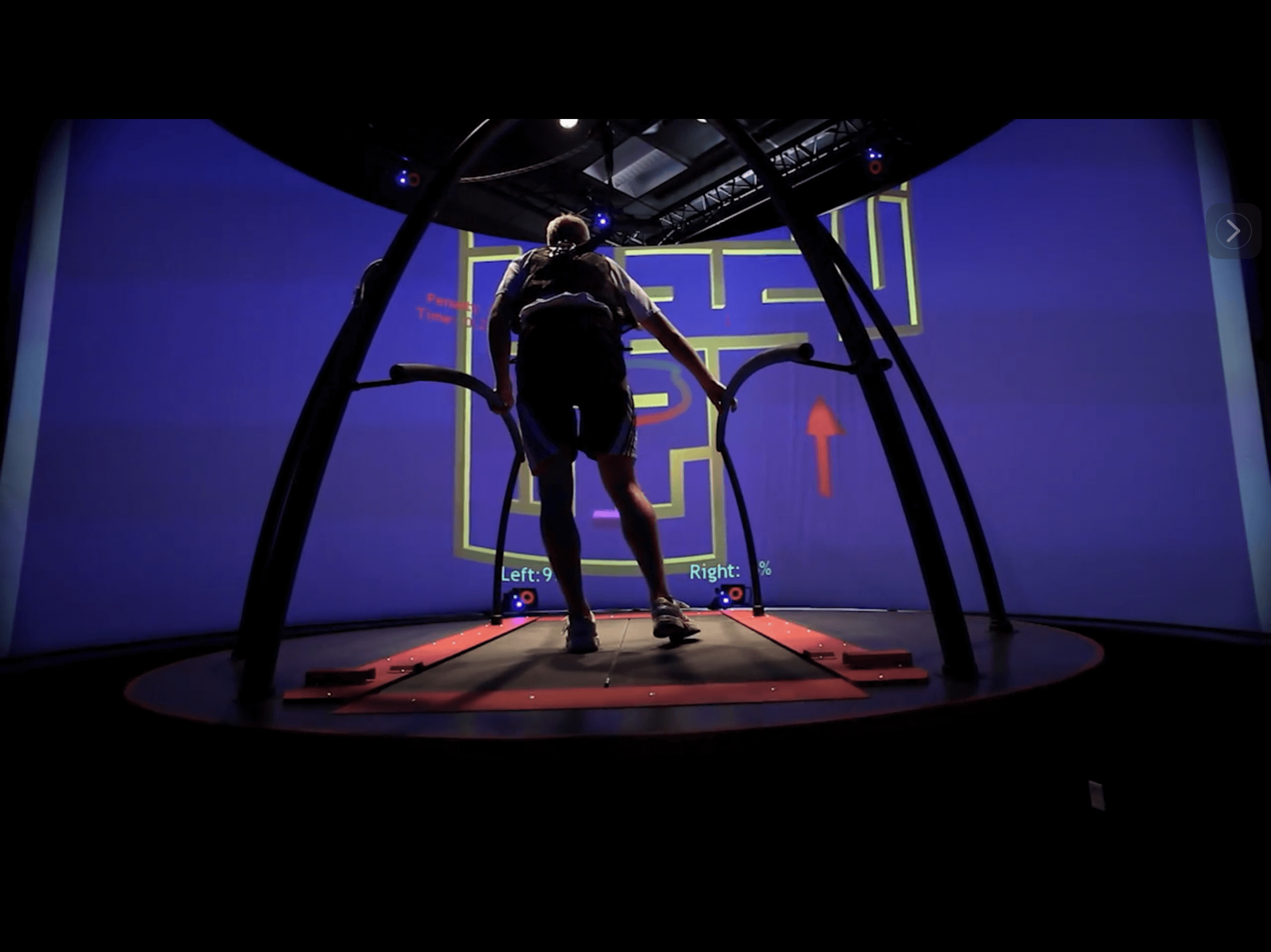
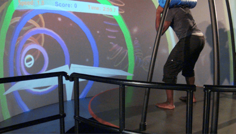
C.A.R.E.N., our computer-assisted rehabilitation environment, provides us with tools to analyze and train patient movement for peak efficiency and optimal execution. The system taps into intrinsic feedback — the use of the patient’s kinesthetic sense that lets them “feel” movement through muscles, joints and the vestibular balance system.
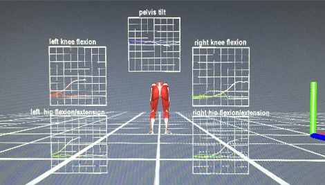
Originally designed to rehab wounded soldiers with multiple traumas, C.A.R.E.N. lets us view joint angles, muscle forces and body symmetry in real time. The combination of sophisticated research grade software and hardware allows multiple feedback possibilities.
With its immersive environment and variety of feedback options, C.A.R.E.N. takes the boredom out of physical therapy while providing the therapist with important real time diagnostic movement data, enabling therapists to achieve goals that would be nearly impossible using tradition overground physical therapy.
In situations where things happen suddenly and unexpected the control through co-contractions is much more important than proprioception. This form of control happens by an instantaneous contraction of all surrounded muscles around the given joint. There is no time delay as there is no feedback of the nervous system needed. It is through the anatomical design and the intrinsic properties that the muscle-tendon units have a direct influence on one and another (all muscles tense and if one lengthens by the perturbation, the other automatically is influenced by it and shortens). This strategy can be trained an optimized over time by the right practice.
Enriched Multi-Sensory Intrinsic Feedback:
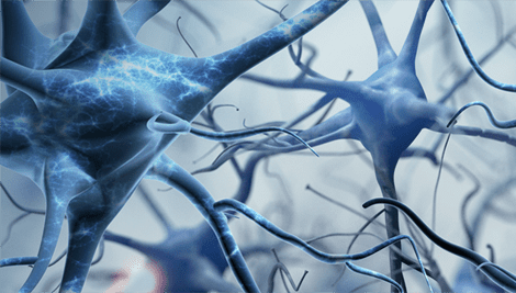
Movement originates in the brain and nervous system. The brain learns best from a dynamic and complex series of experiences, and adapts quickly to environmental stimuli and multi sensory feedback.
Brain Neuroplasticity Potential:
The brain can rewire itself indefinitely in response to imposed stimuli. VR therapy taps into the brain’s neuroplasticity potential to retrain joint movements in response to intrinsic multi-sensory feedback.
Neuroplasticity of the brain is the underlying concept for immersive environment rehabilitation. The brain has the ability to form and reorganize synaptic connections, both functionally and physically in response to new learning or physical therapy following an injury to the neuromusculoskeletal system. Therefore, an individual has the chance to recover faster if the sensory feedback is enhanced. But the brain forms new connections better when the task you are performing has a functional and meaningful goals. Repetitively performing a certain motion or task mindlessly does not help the patient progress or form new synaptic connections in the brain. Research shows that the more time a patient dedicates to therapy with functional goals in mind, the stronger the synapses and faster re-organization of the brain can be achieved.
Immersive Virtual Environment:
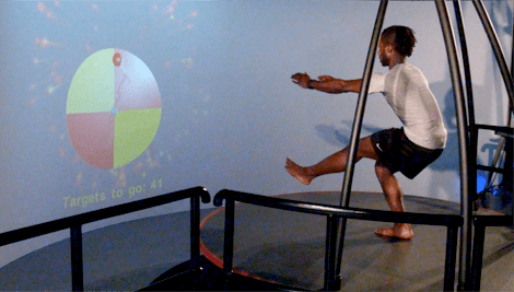
In an immersive VR environment, patients engage and interact in real time with an artificially created environment and its contents. C.A.R.E.N. technology uses sophisticated computer hardware and software to immerse patients and provide feedback about neuromuscular activity. Images, sounds and other stimuli provide an engrossing VR patient experience.
Customized Feedback Training:
C.A.R.E.N. can be programmed to meet the needs of each individual, from an elderly person needing balance training, to an elite athlete recovering from injury. Biomechanical data is presented in real-time to help the patient perform exercises more effectively than with verbal or tactile feedback.
Pain Inhibited via Virtual Feedback:
VR reduces pain perception in otherwise healthy patients suffering chronic low back and knee pain. Fear of pain can make patients reluctant to exercise. Exercise is often key to pain resolution. (em 10/19/18)
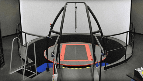
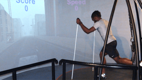
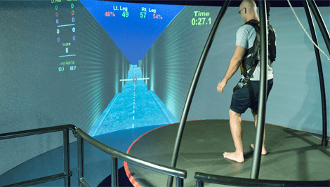
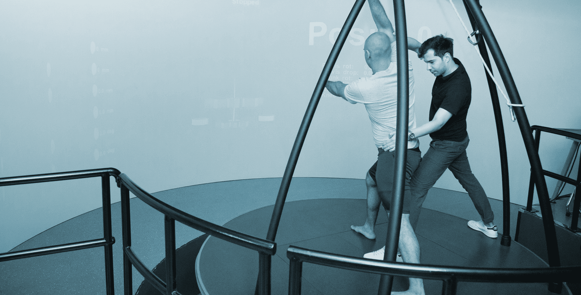
6DOF (six degrees of freedom) Instrumented Platform with Dual Belt Treadmill:
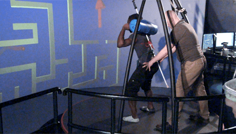
Platform and treadmill instrumentation enables us to mimic and replicate real-life situations in a controlled and protected environment. A safety harness allows the patient to move and interact freely, without fear of falling and injury.
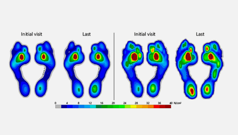
Quantified Ground Reaction Forces and Joint Movements:
Using EMG and force plate technology, therapists can quantify joint angles and measure forces acting on the body. We can then use this data to correct deficiencies and retrain faulty motor patterns.
Identify and Correct Asymmetrical Motor Patterns:
Pain and injury in a joint or limb can lead to compensation patterns during task performance. Compensating can lead to imbalances that create asymmetry. If left uncorrected, asymmetry can lead to further pain and injury. (em 10/21/18)
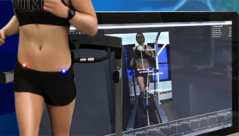
Real Time Muscle and Brain Activity:
C.A.R.E.N. can collect information about muscle and brain activity via EMG or EEG in real time. Athletic injury rehab with C.A.R.E.N. promotes healing and builds confidence. Patients suffering stroke, concussion or neuromuscular disorders can benefit from VR therapy without fear of pain or injury.
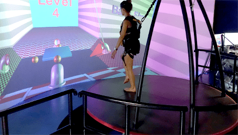
Postural Balance and Stability Training:
Impaired balance and stability pose a threat of serious falls that can lead to injury or even death. C.A.R.E.N is able to assess and improve postural stability and balance control, giving therapists tools for defining baseline measurements, monitoring progress, and individualizing treatment. (em 9/9/18)
VR therapy has been shown to improve health and fitness with reduced pain and discomfort for FM patients. Improve aerobic capacity, coordination, balance and posture. Increase mobility and motivation to exercise in a pain-free VR environment. (em 9/8/18)
A Step Up from Traditional PT:
VR takes the boredom and redundancy out of physical therapy. Instrumentation allows us to adjust the training platform to impose challenges that mimic real-life scenarios. The brain and neuromuscular system adapt in a functional way to VR therapy, preparing the patient for real life experiences.
Dr. Kalika is currently a certified member of:
American Institute of Ultrasound Medicine

Active member of ISMST
International Society of Extra Corporeal Shockwave Therapy
Active member of GCMAS
Gait and Clinical Movement Analysis Society
Active member of NASS
North American Spine Society
Active member of IADMS
International Association of Dance Medicine and Science
Active member of Virtual Rehabilitation Society
Active member of ASRA
American Society of Regional Anesthesia and Pain Medicine
American Academy
Association of Orthopedic Medicine

Active member of Interventional Orthobiologics Foundation


Verified Expert Profiles
Dr. Lev Kalika is a world-recognized expert in musculoskeletal medicine. with 20+ years of clinical experience in diagnostic musculoskeletal ultrasonography, rehabilitative sports medicine and conservative orthopedics. In addition to operating his clinical practice in Manhattan, he regularly publishes peer-reviewed research on ultrasound-guided therapies and procedures. He serves as a peer reviewer for Springer Nature.
Dr. Kalika is an esteemed member of multiple professional organizations, including:
Below is a prime example of how ultrasound can take the guesswork out of diagnosis.

A bad physical therapy experience is one of the primary causes of unnecessary surgery
In this instance, an athlete was originally diagnosed with minor quadriceps muscle strain and was treated for four weeks, with unsatisfactory results. When he came to our clinic, the muscle was not healing, and the patients’ muscle tissue had already begun to atrophy.
Upon examination using MSUS, we discovered that he had a full muscle thickness tear that had been overlooked by his previous provider. To mitigate damage and promote healing, surgery should have been performed immediately after the injury occurred. Because of misdiagnosis and inappropriate treatment, the patient now has permanent damage that cannot be corrected.
The most important advantage of Ultrasound over MRI imaging is its ability to zero in on the symptomatic region and obtain imaging, with active participation and feedback from the patient. Using dynamic MSUS, we can see what happens when patients contract their muscles, something that cannot be done with MRI. From a diagnostic perspective, this interaction is invaluable.
Dynamic ultrasonography examination demonstrating
the full thickness tear and already occurring muscle atrophy
due to misdiagnosis and not referring the patient
to proper diagnostic workup
Demonstration of how very small muscle defect is made and revealed
to be a complete tear with muscle contraction
under diagnostic sonography (not possible with MRI)

Complete tear of rectus femoris
with large hematoma (blood)

Separation of muscle ends due to tear elicited
on dynamic sonography examination


