Your thoracic spine plays an important role in conducting nerve impulses and mediating load transfers during physical activity. When the muscles and fascia that affect your mid-back are overly weak, tight or imbalanced, your spine becomes unstable and mobility is reduced, putting you at risk for pain and injury. Accurate diagnosis and advanced treatment technologies are game-changers when it comes to middle back pain treatment in NYC.
or
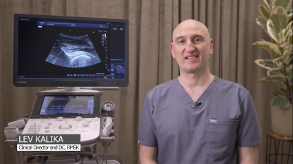
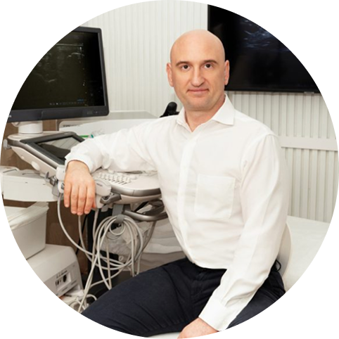
Clinical director & DC RMSK
Verified Expert Profiles
Dr. Kalika , NYDNRehab clinical director, is an internationally recognized expert in the field of human movement disorders, with multiple scientific publications to his credit. His research topics include:
Dr. Kalika is certified in multiple evidence-based therapy approaches. His clinic boasts the most advanced technologies available for treating pain syndromes and movement disorders. Such advanced technologies are found in only a small fraction of private clinics, and are rarely available to the general public.
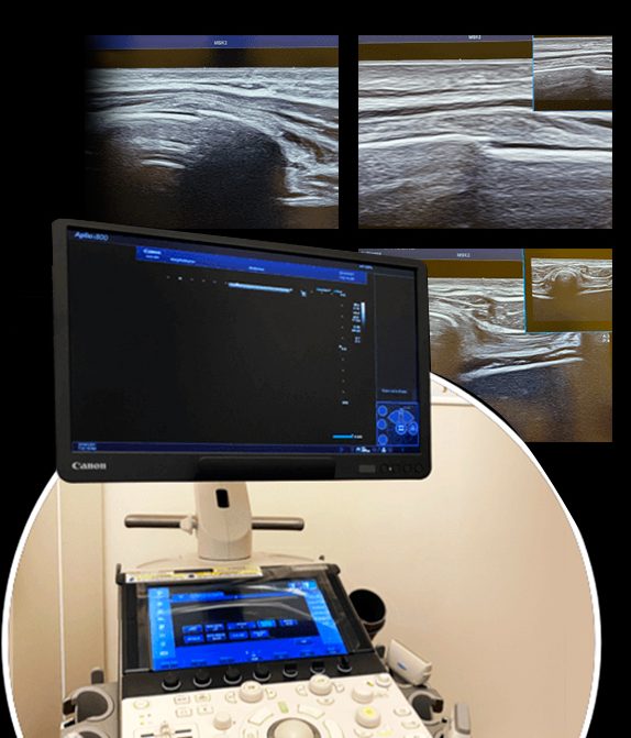
Dr. Kalika specializes in high resolution diagnostic musculoskeletal ultrasonography. Ultrasound imaging enables us to detect middle back conditions such as scapular dyskinesis and issues affecting the nerves of the trunk, which are the most frequent causes of middle back pain.
Dr. Kalika has over 20 years of experience in DNS therapy, the thoracic ring approach, shockwave therapy (ESWT), and ultrasound guided dry needling. His experience and expertise make him one of the world’s most versatile practitioners for mid-back pain and thoracic spinal disorders. Hundreds of patients rely on Dr. Kalika for the best middle back pain chiropractic treatment.
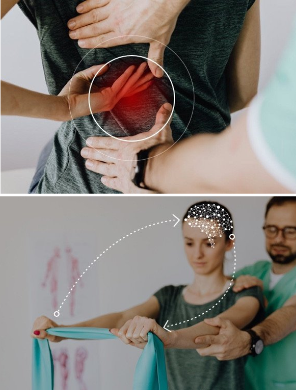
Physical therapy is a valuable and effective approach to resolving musculoskeletal pain and dysfunction, but in many cases, physical therapy does not provide a stand-alone solution. Prior to beginning physical therapy, patients often need to address underlying issues that contribute to their pain and disability.
Unfortunately, mainstream physical therapy clinics are often not adequately equipped or experienced to identify and treat complications that undermine the effectiveness of physical therapy. They often rely on one-size-fits-all treatment protocols that overlook the unique characteristics of the individual condition, opting to treat the symptoms and not the patient.
Identifying and treating underlying issues prior to beginning physical therapy is key to getting fast and effective results. Failure to do so can completely undermine your treatment protocol, and in some cases, your condition may even worsen.
At NYDNRehab, we use a broad range of advanced technologies and innovative therapeutic approaches to resolve issues that can potentially undermine the success of physical therapy.
Our talented staff is certified in a diverse array of treatment methodologies, rarely found in run-of-the-mill physical therapy clinics. Our one-on-one sessions are personalized, based on the patient’s unique diagnostic profile.
At NYDNRehab, we pride ourselves on providing the best possible care for our valued patients. Our personalized treatment protocols and one-on-one approach ensure that you get the exact therapy you need to resolve your middle back pain and dysfunction.
Run-of-the-mill clinics often take a one-size-fits-all approach to middle back pain physical therapy, overlooking fundamental differences between one patient and the next. We custom-design our treatment protocols based on your diagnostic results, to ensure that you receive the very best personalized care for your mid-back pain.
Our growing arsenal of high-tech tools guarantee that you will get a fast and accurate diagnosis. Our cutting-edge treatment methodologies and advanced technologies ensure that your pain and dysfunction are successfully resolved in the shortest time possible.
Our approach, technologies, experience and expertise make NYDNRehab the clinic of choice for middle back pain treatment in NYC.
Fractured or bruised ribs
Shoulder blade instability
Scoliosis
Degenerative disc disease
Poor posture
Herniated disc
Arthritis
Osteoporosis
Obesity
Muscle weakness and imbalances
Poor flexibility
Your middle back houses a complex network of bones, muscles, nerves and connective tissues that work interdependently to stabilize your torso and produce movement. Pain and dysfunction in the middle back region can have multiple causes, and symptoms alone do not provide enough information to arrive at an accurate diagnosis.
At NYDNRehab, we use the highest resolution ultrasound imaging to visualize the structures of the middle back in real time, with the patient in motion. The flexibility of ultrasonography makes it superior to MRI or Xray as a diagnostic tool. Our ultra-sensitive high-tech transducers provide clarity, depth and detail, so we don’t miss a thing.
Your thoracic spine is the longest and most complex area of your spinal column, made up of 12 vertebrae that span between your cervical and lumbar spinal regions. The bones that make up your rib cage attach to the thoracic vertebrae, making the mid-back an important component of your breathing apparatus.
In addition, movement in your thoracic region is governed by a complex network of muscles and connective tissues that play a major role in sports and physical activity.
Instability, reduced mobility or injury to the middle back can affect your breathing, impair your movement and increase your risk of injury during physical activity.
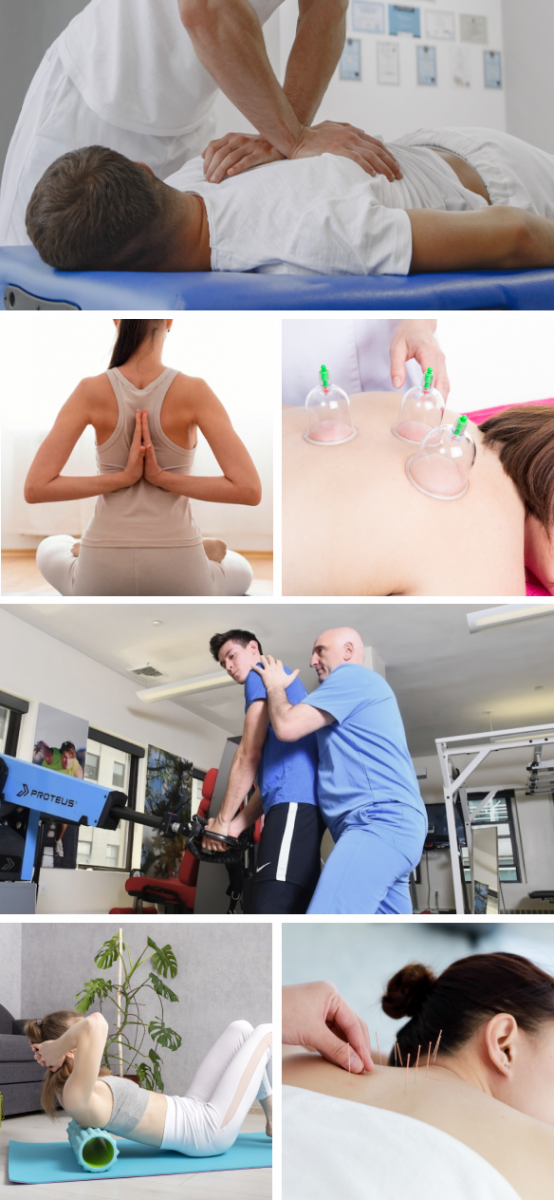
Your personalized treatment protocol will depend on the exact nature of your mid-back issues. Your middle back pain treatment may include some or all of the following therapies:
The complexity of the mid-back region and shoulder girdle makes it difficult to identify the exact source of pain and dysfunction. In many cases, multiple factors come into play. High resolution ultrasound imaging is an advanced and effective diagnostic tool, especially when coupled with new technologies that dramatically enhance visualization and facilitate rehabilitation.
ShowMotion is an objective tool for joint movement analysis that uses motion tracking sensors, placed on the patient’s skin to collect data about movement quality. The patient performs a series of joint-specific movements, and the data is analyzed by ShowMotion’s proprietary software and displayed on a computer screen. The collected information provides valuable insights about inefficient movement patterns, compensation patterns, and improvements in movement in response to therapy, allowing for personalized rehabilitation.
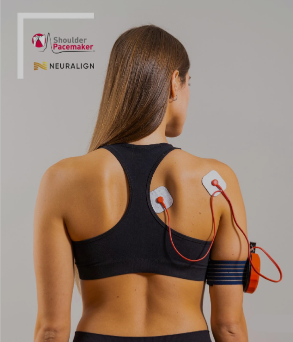
The Neuralign Shoulder Pacemaker is a shoulder rehabilitation device with a kinematic sensor activated by movement. The patient dynamically interacts with the device to stimulate efficient muscle recruitment patterns, enhance movement quality, and restore optimal muscle balance during rehabilitation. The sensor provides objective data that practitioners can use to support decision-making and personalize shoulder rehabilitation.
The Shoulder Pacemaker can be used to target several common conditions:
The Shoulder Pacemaker is also a cutting-edge tool for enhancing athletic performance in shoulder-intensive sports like swimming, tennis, baseball and others.
Your body has its own innate healing mechanisms, but they sometimes need a nudge to accelerate the healing process. Regenerative technologies help to jump-start healing by stimulating tissue repair at the cellular level, with minimal discomfort for the patient.
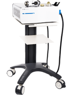
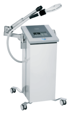
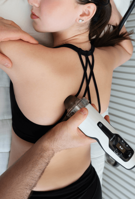
Focused ESWT is a regenerative treatment for damaged tendon, muscle and bone tissue. This technology produces high frequency sound waves to stimulate the body’s own reparative mechanisms.
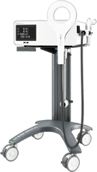
EMTT is a fairly new technology that transmits high energy magnetic pulses to targeted tissues. The magnetic waves synchronize with the body’s own magnetic fields to trigger a regenerative response. EMTT waves can penetrate deep tissues, to target difficult-to-reach tendons, muscles, bones and nerves.
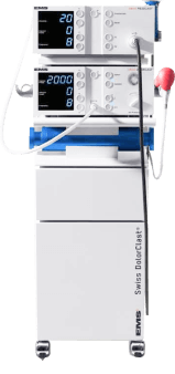
Extracorporeal Pulse Activation Technology (EPAT)
EPAT, also known as defocused shock wave therapy, uses acoustic pressure waves to enhance blood circulation to targeted tissues, to accelerate the healing process.
HEIT uses electromagnetic fields to penetrate cells, tissues, organs and bones, to reactivate the electrochemical function of cells and cell membranes.
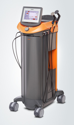
Our INDIBA Tecar therapy machine converts electrical current into a stable radio frequency current of 448 kHz, designed to increase and stabilize the exchange of ions in damaged cells, evoking a regenerative response that accelerates healing. INDIBA can be used to successfully treat joint and muscle disorders, low-back pain, sports injuries, surgical incisions and various pain syndromes. Another therapeutic effect of INDIBA is extreme and prolonged cellular hyperthermia. Due to this effect, INDIBA therapy combined with manual therapy and soft tissue tissue manipulation enables instantaneous release to occur, significantly shortening the number and duration of physical therapy sessions. What is normally accomplished in two months of physical therapy can be accomplished in 3-4 sessions with INDIBA.
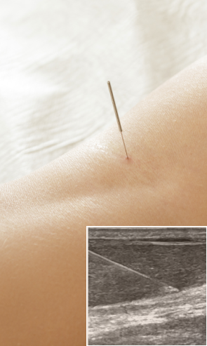
Myofascial trigger points often contribute to lower back pain. Dry needling is an outpatient procedure that inserts non-medicated needles into the trigger point to evoke a twitch response, releasing the trigger point and immediately relieving pain. Ultrasound guidance eliminates the need for multiple insertions, reducing pain and discomfort for the patient.
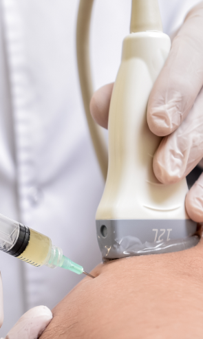
Injection therapies use natural/neutral solutions that stimulate cellular repair by either nourishing or irritating the targeted cells. Guidance by ultrasound ensures that the injected substances hit their mark, for maximum effectiveness.
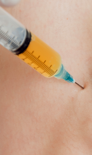
PRP therapy uses a sample of the patient’s own whole blood, which is spun in a centrifuge to extract a high concentration of platelets. When injected into damaged tissues, PRP initiates tissue repair by releasing biologically active factors such as growth factors, cytokines, lysosomes and adhesion proteins. The injected solution stimulates the synthesis of new connective tissues and blood vessels. PRP can help to jump-start healing in chronic injuries and accelerate repair in acute injuries.
Prolotherapy uses a biologically neutral solution, often containing dextrose, saline or lidocaine (Read More). The solution irritates the affected connective tissue, stimulating the body’s own natural healing mechanisms to encourage growth of new normal ligament or tendon fibers.
These case studies reflect real clinical conditions evaluated at NYDNRehab using advanced diagnostic methods and individualized rehabilitation strategies. All cases are evaluated and managed by Dr. Lev Kalika and the NYDNRehab clinical team.
Middle back pain and dysfunction can lead to major disability over time, increasing your risk of injuries and severely impacting your quality of life. You don’t have to live with mid-back pain. With a growing arsenal of technological tools at our disposal, the back pain specialists at NYDNRehab are able to accurately diagnose and successfully treat middle back pain and dysfunction without dangerous drugs or surgery. Contact NYDNRehab today, and get the middle back pain treatment you need so you can get back to doing the things you love.


Verified Expert Profiles
Dr. Lev Kalika is a world-recognized expert in musculoskeletal medicine. with 20+ years of clinical experience in diagnostic musculoskeletal ultrasonography, rehabilitative sports medicine and conservative orthopedics. In addition to operating his clinical practice in Manhattan, he regularly publishes peer-reviewed research on ultrasound-guided therapies and procedures. He serves as a peer reviewer for Springer Nature.
Dr. Kalika is an esteemed member of multiple professional organizations, including:Independent peer-reviewed research relevant to this treatment approach.
Research authored or co-authored by the clinic’s medical director. The following research publications inform the clinical approach used in this treatment program.
Conference abstract
2025
 Lev Kalika
  Rostyslav Bubnov
 Conference abstract
2025
 Lev Kalika Rostyslav Bubnov
Below is a prime example of how ultrasound can take the guesswork out of diagnosis.

A bad physical therapy experience is one of the primary causes of unnecessary surgery
In this instance, an athlete was originally diagnosed with minor quadriceps muscle strain and was treated for four weeks, with unsatisfactory results. When he came to our clinic, the muscle was not healing, and the patients’ muscle tissue had already begun to atrophy.
Upon examination using MSUS, we discovered that he had a full muscle thickness tear that had been overlooked by his previous provider. To mitigate damage and promote healing, surgery should have been performed immediately after the injury occurred. Because of misdiagnosis and inappropriate treatment, the patient now has permanent damage that cannot be corrected.
The most important advantage of Ultrasound over MRI imaging is its ability to zero in on the symptomatic region and obtain imaging, with active participation and feedback from the patient. Using dynamic MSUS, we can see what happens when patients contract their muscles, something that cannot be done with MRI. From a diagnostic perspective, this interaction is invaluable.
Dynamic ultrasonography examination demonstrating
the full thickness tear and already occurring muscle atrophy
due to misdiagnosis and not referring the patient
to proper diagnostic workup
Demonstration of how very small muscle defect is made and revealed
to be a complete tear with muscle contraction
under diagnostic sonography (not possible with MRI)

Complete tear of rectus femoris
with large hematoma (blood)

Separation of muscle ends due to tear elicited
on dynamic sonography examination