During pregnancy, the elasticity of your abdominal muscles enables them to stretch to accommodate your growing baby. However, the linea alba is made up mostly of collagen, which is less elastic than muscle. For many women, the linea alba thins during pregnancy and gradually tightens up again over several weeks postpartum. But in some instances, the linea alba is stretched to the point of separation, leaving an undesirable gap between the right and left sides of the RA.
Not only is diastasis recti cosmetically undesirable, giving you a postpartum tummy that will not go away, but it can interfere with the overall function of your abdominal canister, leading to a plethora of related issues.or
Clinical director & DC RMSK
Verified Expert Profiles
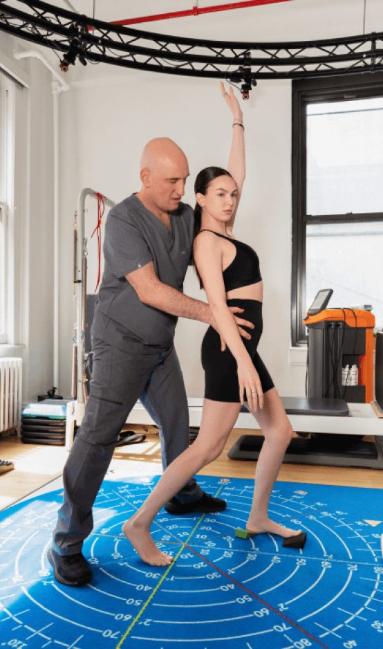
The pelvic pain specialists at NYDNR use a targeted conservative approach to treating diastasis recti. Our therapy is individualized, taking into account the specific needs of each patient.
Physical therapy is a valuable and effective approach to resolving musculoskeletal pain and dysfunction, but in many cases, physical therapy does not provide a stand-alone solution. Prior to beginning physical therapy, patients often need to address underlying issues that contribute to their pain and disability.
Unfortunately, mainstream physical therapy clinics are often not adequately equipped or experienced to identify and treat complications that undermine the effectiveness of physical therapy. They often rely on one-size-fits-all treatment protocols that overlook the unique characteristics of the individual condition, opting to treat the symptoms and not the patient.
Identifying and treating underlying issues prior to beginning physical therapy is key to getting fast and effective results. Failure to do so can completely undermine your treatment protocol, and in some cases, your condition may even worsen.
At NYDNRehab, we use a broad range of advanced technologies and innovative therapeutic approaches to resolve issues that can potentially undermine the success of physical therapy. Our talented staff is certified in a diverse array of treatment methodologies, rarely found in run-of-the-mill physical therapy clinics. Our one-on-one sessions are personalized, based on the patient’s unique diagnostic profile.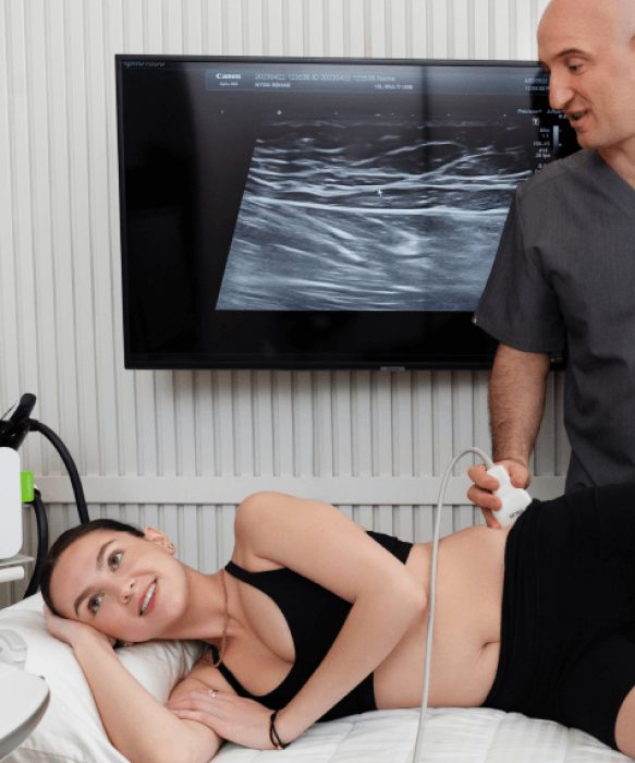
Getting in shape before pregnancy, maintaining a healthy weight and practicing lifestyle behaviors that promote healthy collagen production can reduce your risk of diastasis recti.

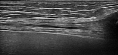
The best and the most precise way to assess diastasis recti is by using diagnostic ultrasonography. The benefits are:

In an effort to flatten a postpartum belly, many women begin an exercise program of challenging abdominal exercises like crunches and leg lifts, but those exercises can sometimes make the condition worse. Other women try wrapping the abdomen to draw the RA together, hoping the collagen will repair itself, which is ineffective. In severe cases, some women even have cosmetic surgery to close the gap. However, cosmetic solutions do not restore full function to the abdominal canister, which is the real problem posed by diastasis recti. Diastasis recti physical therapy offers a conservative, non-invasive treatment solution.
Your body has its own innate healing mechanisms, but it sometimes needs a nudge to accelerate the healing process. Non-invasive regenerative technologies help to jump-start healing by stimulating tissue repair at the cellular level.
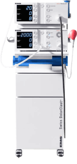
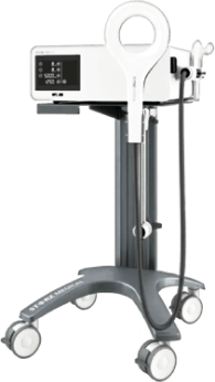
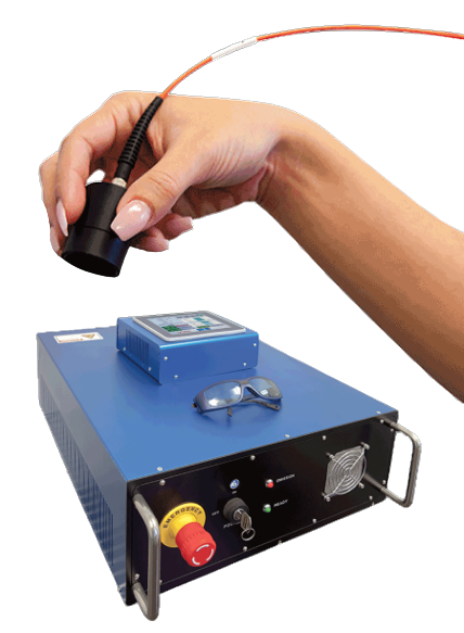
Focused Shockwave Therapy
Focused ESWT technology produces high frequency sound waves to reduce pain and inflammation and stimulate the body’s own reparative mechanisms.
Our new PiezoWave shockwave machine features MyACT, a new type of focused shockwave technology that allows for deeper compression of the focused waves. Its higher frequency allows for precise neuro modulation under ultrasound guidance, with a special linear head for treating myofascial pain.
This innovative shockwave technology transforms the mechanical energy of shockwaves into biochemical signals that precisely target damaged tissues. Its versatile system of controls lets us fine-tune its output levels for precise and personalized patient care. With MyACT, we are able to repair and regenerate damaged tissues by stimulating the body’s own reparative mechanisms.
Myofascial Acoustic Compression Therapy (MyACT)
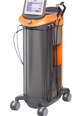
Extracorporeal Pulse Activation Technology (EPAT)
EPAT, also called radial shock wave therapy, is not a true shockwave, but uses mechanical pressure waves that are much weaker than focused shockwaves. EPAT is most useful in regenerating fascia fibroblasts and eliminating fascia densifications.
EMTT transmits high energy magnetic pulses that synchronize with your body’s own magnetic fields, triggering a regenerative response in tendons, muscles, bones and nerves.
Extracorporeal Magnetic Transduction Therapy (EMTT)
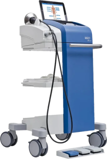
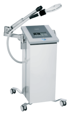
INDIBA Radiofrequency Therapy
INDIBA therapy helps to restore the ionic charge of damaged cells, for faster injury healing.
The NESA device generates a biphasic low-frequency electrical current that travels through autonomic neural pathways, to restore optimal neural signaling to the brain.
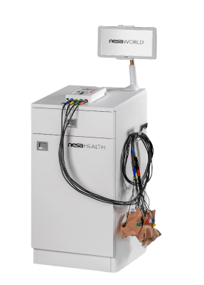
NESA Neuromodulation Therapy
HIGH ENERGY INDUCTIVE THERAPY (HEIT)
High Energy Inductive Therapy (HEIT) is a new technology that uses electromagnetic fields to penetrate cells, tissues, organs and bones, to reactivate the electrochemical function of cells and cell membranes. HEIT relieves pain by restoring peripheral nerve function.
Laser therapy is a non-invasive approach to treating chronic pain that reduces inflammation, eliminates pain and speeds up tissue healing. The treatment uses light energy to penetrate your skin, muscle, nerve, and connective tissues, stimulating a photochemical reaction that changes the chemical and physical properties of the affected area at the molecular level
Phoenix Theralase Laser Therapy
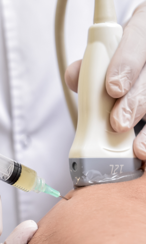
Injection therapies use natural/neutral solutions that stimulate cellular repair by either nourishing or irritating the targeted cells. Guidance by ultrasound ensures that the injected substances hit their mark, for maximum effectiveness.
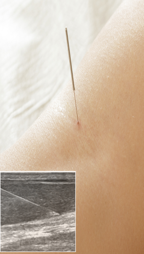
Myofascial trigger points often contribute to lower back pain. Dry needling is an outpatient procedure that inserts non-medicated needles into the trigger point to evoke a twitch response, releasing the trigger point and immediately relieving pain. Ultrasound guidance eliminates the need for multiple insertions, reducing pain and discomfort for the patient.
Hydrodissection is a technique for treating peripheral nerve entrapment. It uses a saline solution to separate entrapped nerves from surrounding fascia adhesions or adjacent structures.
The pelvic pain specialists at NYDNR use a targeted conservative approach to treating diastasis recti. Our therapy is individualized, taking into account the specific needs of each patient.


These case studies reflect real clinical conditions evaluated at NYDNRehab using advanced diagnostic methods and individualized rehabilitation strategies. All cases are evaluated and managed by Dr. Lev Kalika and the NYDNRehab clinical team.

Below is a prime example of how ultrasound can take the guesswork out of diagnosis.

A bad physical therapy experience is one of the primary causes of unnecessary surgery
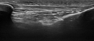
In this instance, an athlete was originally diagnosed with minor quadriceps muscle strain and was treated for four weeks, with unsatisfactory results. When he came to our clinic, the muscle was not healing, and the patients’ muscle tissue had already begun to atrophy.
Upon examination using MSUS, we discovered that he had a full muscle thickness tear that had been overlooked by his previous provider. To mitigate damage and promote healing, surgery should have been performed immediately after the injury occurred. Because of misdiagnosis and inappropriate treatment, the patient now has permanent damage that cannot be corrected.
The most important advantage of Ultrasound over MRI imaging is its ability to zero in on the symptomatic region and obtain imaging, with active participation and feedback from the patient. Using dynamic MSUS, we can see what happens when patients contract their muscles, something that cannot be done with MRI. From a diagnostic perspective, this interaction is invaluable.
Dynamic ultrasonography examination demonstrating
the full thickness tear and already occurring muscle atrophy
due to misdiagnosis and not referring the patient
to proper diagnostic workup
Demonstration of how very small muscle defect is made and revealed
to be a complete tear with muscle contraction
under diagnostic sonography (not possible with MRI)

Complete tear of rectus femoris
with large hematoma (blood)

Separation of muscle ends due to tear elicited
on dynamic sonography examination