Clinical director & DC RMSK
Verified Expert Profiles

Orthobiologic, Sports Medicine and Regenerative Medicine Specialist
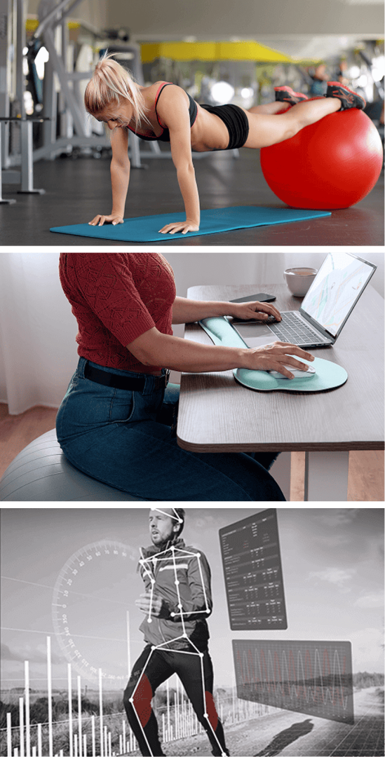
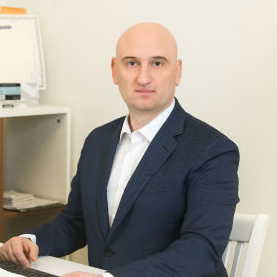
Sedentary lifestyle and excessive sitting
Being overweight or obese
Weak core and pelvic muscles
Weak and imbalanced spinal stabilizers
Fascial dysfunction
Poor posture
Deficits in gait mechanics
Inefficient lifting technique
Repetitive overuse from sports, exercise or occupation
Old injuries that were never properly rehabilitated
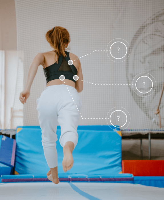
It is commonly thought that low back pain begins in the spine, but in reality, while symptoms are felt in the lower spine, the problem often starts elsewhere. Unlike conventional medical doctors who focus on treating the spine itself, holistic practitioners seek to identify and correct the causes of low back pain, with the goal of restoring pain-free mobility.
The lumbar spine and pelvis are primary sites of load transfer between the upper and lower body, and the spine houses sensitive neural bodies that respond to movement, posture, and mechanical stress. Inefficient gait mechanics, poor load distribution, fascial restrictions, and nerve irritation often originate in the lower kinetic chain, with the lower back absorbing the consequences.
Treatment centered on the spine may provide temporary pain relief, but the symptoms are likely to return if other issues are not addressed. Our integrative approach takes into account biomechanical factors, lifestyle behaviors, inefficient motor strategies, and postural habits. By identifying multiple factors that contribute to the patient’s low back pain, we can design a customized plan to treat and correct them, without drugs or surgery.
Benefits of holistic vs conventional LBP treatment include:
Many people assume that lower back pain begins and ends in the spine. In reality, the lower back is often where symptoms appear — not where the problem starts. The lumbar spine functions as a central load-transfer zone between the feet, hips, pelvis, and upper body, while also housing sensitive neural structures that respond to movement, posture, and mechanical stress.
When inefficient gait mechanics, poor load distribution, fascial restrictions, or nerve irritation exist elsewhere in the kinetic chain, the lower back often absorbs the consequences. In these cases, treatments focused only on the spine may provide temporary relief, while symptoms return once normal activity resumes.
This is why persistent or recurring low back pain often requires evaluating how the body moves, loads, and adapts — not just how the spine looks at rest.
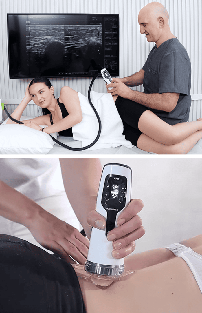
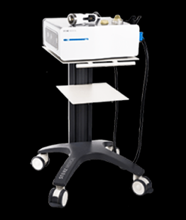
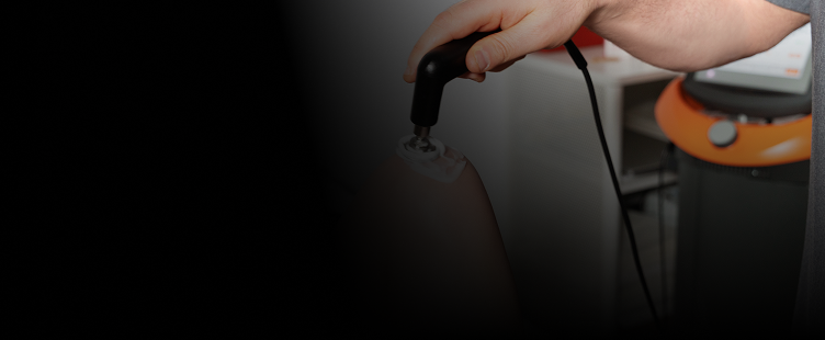
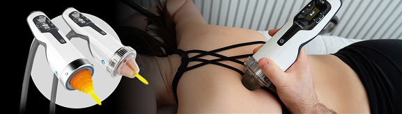
Extracorporeal shockwave therapy (ESWT) delivers high-energy acoustic waves to targeted tissues. At NYDNRehab, we use a combination of focal, radial, linear and defocused shockwaves, based on the type, depth and area of damaged tissue. The waves induce mechanotransduction, converting mechanical energy into biochemical signals that promote cellular repair.
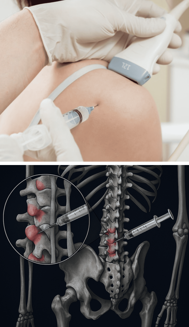
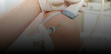
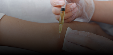
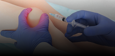
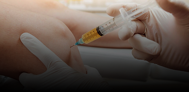
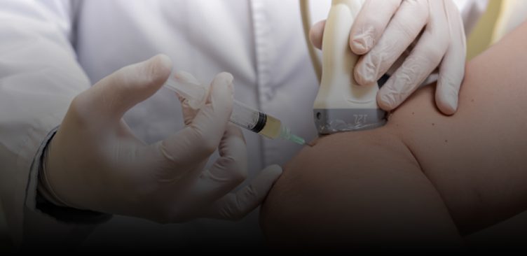
At NYDNRehab, we guide our Prolotherapy procedures with high-resolution ultrasound, to ensure the solution precisely reaches its target. Injections performed without imaging often miss their mark, and can bleed into adjacent tissues, reducing their efficacy. When combined, Prolotherapy and ESWT can dramatically reduce low back pain and set the patient up for physical therapy success.
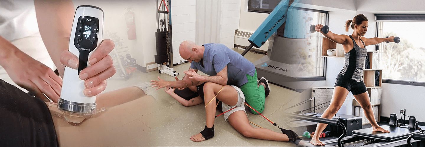
Regenerative therapies and orthobiologic procedures work together to stimulate and speed up tissue healing while dramatically reducing pain and inflammation. Pre-treatment with these cutting-edge therapies sets you up for physical therapy success and lasting results.
We guide our procedures with high-resolution ultrasonography, to maximize their effectiveness. Your treatment plan may include a combination of therapies, based on your unique condition.
Regenerative therapies and orthobiologic procedures are often administered as stand-alone solutions to treat damaged tissues and pain syndromes. But while that approach may provide temporary relief, it rarely resolves the problem over the long term. By carefully selecting and combining the most appropriate therapies, we are able to harness the synergistic effects of both regenerative therapies and orthobiologics, for superior and enduring results.
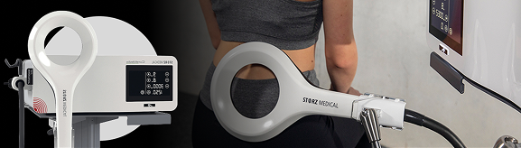
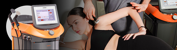
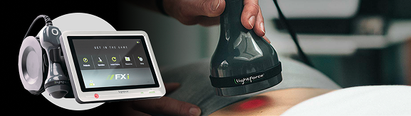
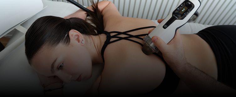
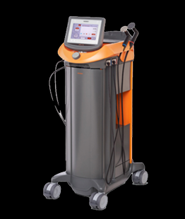
When delivered at the most effective levels under ultrasound guidance, ESWT + EMTT prime the microenvironment via mechanical and electromagnetic stimulation. TECAR + Laser therapy optimizes tissue metabolism, circulation, and cellular energy. Together, they extend and amplify the biological effects of PRP and stem cells, accelerating tissue integration, repair, and functional recovery.
In addition, when used post-procedure, regenerative therapies dramatically reduce pain and discomfort, reinforce soft tissue healing, and promote collagen fiber realignment.
Despite the effectiveness of these cutting-edge therapies, without a multifaceted approach that addresses not only symptoms and tissue healing, but also causative and biomechanical factors, the patient is unlikely to restore and maintain efficient pain-free mobility over the long term. At NYDNRehab, our goal is to not only address symptoms and heal tissues, but to optimize mobility and prevent future injuries.
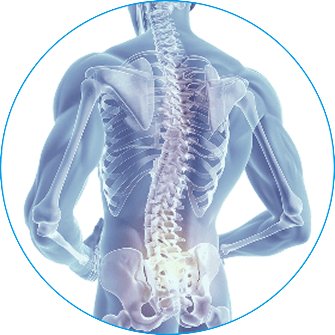
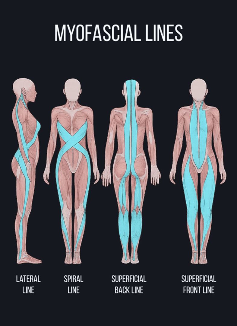
The lower back is supported by strong ligaments, muscles, and fascia that hold the spine in alignment, control movement, and distribute forces. Healthy, fascia is smooth, slippery and elastic, enabling muscles, nerves and blood vessels to glide among other structures. But damaged fascia can become dense and sticky, inhibiting coordinated movement, and compressing nerves, which generates pain signals.
The Stecco technique of fascial manipulation targets densified fascial layers, helping to restore their gliding and elastic properties. Stecco Fascial Manipulation is a highly systematic approach that requires specific training. When applied correctly, Stecco not only restores tissue gliding – it reactivates sensory receptors within the fascia, recalibrating the neuromuscular system and restoring efficient motor control.
Progression of Stecco Fascial Manipulation involves:
This advanced methodology works to alleviate lower back pain, eliminate restricted movement, enhance physical performance, and optimize whole-body mobility and stability. Dr. Kalika has received advanced training in the Stecco Method directly from its creator, Dr. Carla Stecco, the world’s leading specialist in fascial science.
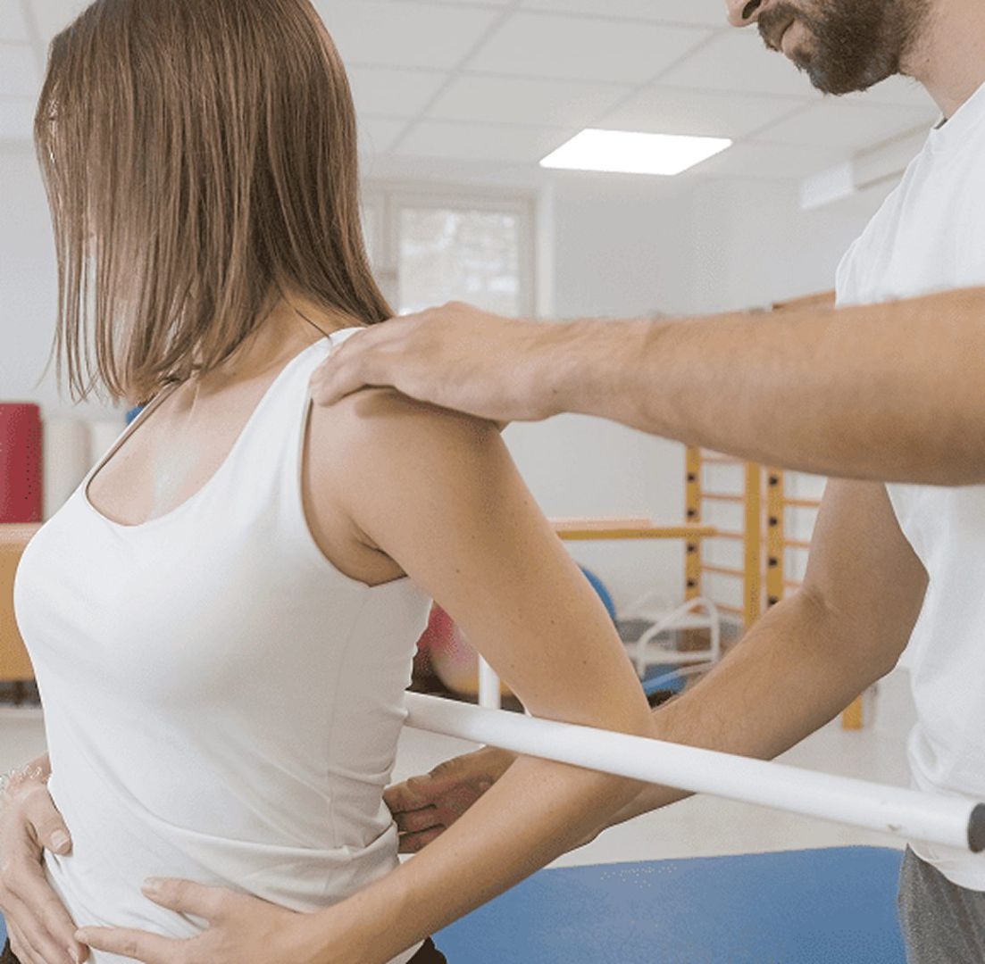
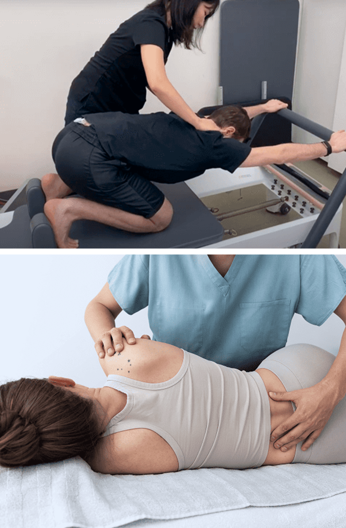
All human movement affects and is affected by the spine, and good spinal alignment is fundamental to overall health. Holistic physical therapy for low back pain seeks to optimize spinal mobility and stability by addressing factors that disrupt alignment, compress nerves, and alter muscle recruitment patterns.
Prior to beginning physical therapy, we pre-treat your tissues to reduce pain and inflammation and promote healing. It is not uncommon for medical doctors to recommend physical therapy after managing pain with medications, but that approach can do more harm than good if the tissues remain damaged. At NYDNRehab, we make sure the structures surrounding and supporting your lower back are ready to handle loads, without pain or risk of injury.
Mainstream approaches to back pain treatment often involve pain medications, muscle relaxants, steroid injections, and admonishments to “take it easy.” When those methods fail to alleviate pain and dysfunction, surgery is often the next step. However, research shows that conservative care can be just as effective as surgery, without the risk and expense of unnecessary invasive procedures.
At NYDNRehab, we work with you to resolve the underlying causes of lower back pain. Our personalized one-on-one approach ensures that you get the very best treatment for your unique condition. If you are ready to eliminate lower back pain for good, contact us today, and get back to doing the things you love.
These case studies reflect real clinical conditions evaluated at NYDNRehab using advanced diagnostic methods and individualized rehabilitation strategies. All cases are evaluated and managed by Dr. Lev Kalika and the NYDNRehab clinical team.
Traditional approaches to back pain treatment often involve pain medications, muscle relaxants, steroid injections, and admonishments to “take it easy.” When those methods fail to alleviate pain and dysfunction, surgery is often the next step. However, research shows that conservative care is at least as effective as surgery, without the risk and expense of unnecessary invasive procedures.
At NYDNRehab, we work with you to resolve the underlying causes of lower back pain. Our personalized one-on-one approach ensures that you get the very best treatment for your unique condition. If you are ready to eliminate lower back pain for good, schedule a consultation with our lower back pain specialists today, and get back to doing the things you love, pain-free!
Clinical director & DC RMSK


 Dr. Yuri Brosgol
MD
Dr. Yuri Brosgol
MD
 Dr. Michael Goynatsky
DPT
Dr. Michael Goynatsky
DPT
 Dr. Daniela Escudero
DPT
Dr. Daniela Escudero
DPT
 Dr. Michelle Agyakwah
DC
Dr. Michelle Agyakwah
DC
 Dr. Tatyana Kapustina
L. Ac.
Dr. Tatyana Kapustina
L. Ac.
Independent peer-reviewed research relevant to this treatment approach.
Research authored or co-authored by the clinic’s medical director. The following research publications inform the clinical approach used in this treatment program.
Conference Abstract
2022
 Lev Kalika
  Rostyslav Bubnov
 Conference Article / Abstract
2018
Lev Kalika Rostyslav Bubnov Conference Abstract
2023
Lev Kalika Rostyslav Bubnov Conference Abstract
2023
Lev Kalika Rostyslav Bubnov Conference Article / Abstract
2018
Lev Kalika Rostyslav Bubnov Conference E‑Poster
2025
Rostyslav Bubnov Lev Kalika Conference E‑Poster
2025
Rostyslav Bubnov Lev Kalika Conference Abstract
2025
Lev Kalika Rostyslav Bubnov 
Below is a prime example of how ultrasound can take the guesswork out of diagnosis.

A bad physical therapy experience is one of the primary causes of unnecessary surgery
In this instance, an athlete was originally diagnosed with minor quadriceps muscle strain and was treated for four weeks, with unsatisfactory results. When he came to our clinic, the muscle was not healing, and the patients’ muscle tissue had already begun to atrophy.
Upon examination using MSUS, we discovered that he had a full muscle thickness tear that had been overlooked by his previous provider. To mitigate damage and promote healing, surgery should have been performed immediately after the injury occurred. Because of misdiagnosis and inappropriate treatment, the patient now has permanent damage that cannot be corrected.
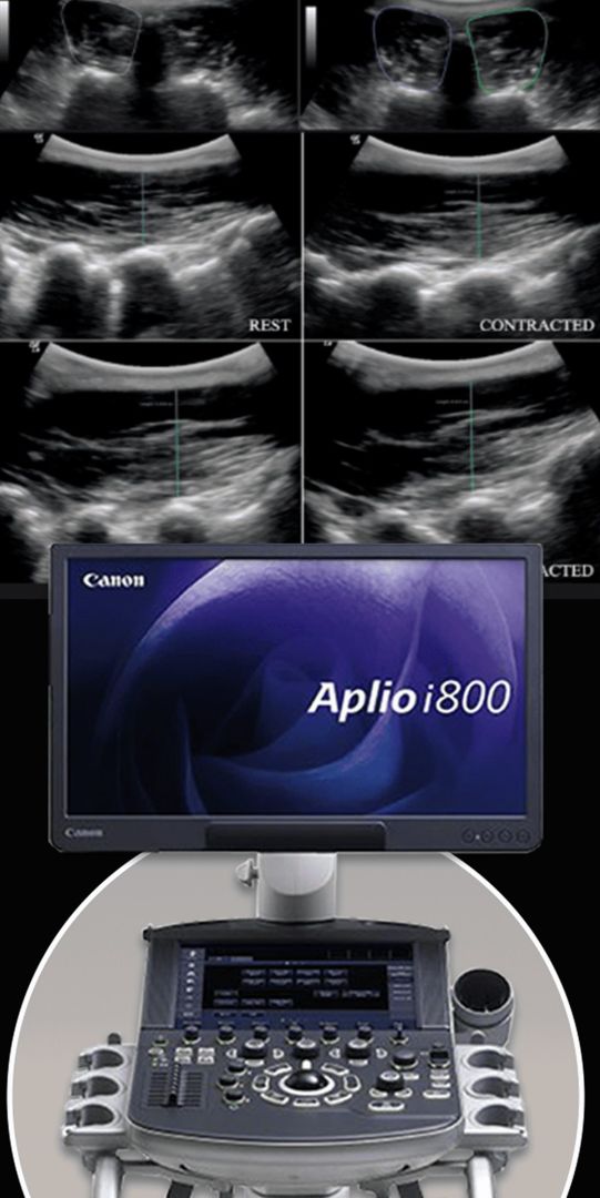
The most important advantage of Ultrasound over MRI imaging is its ability to zero in on the symptomatic region and obtain imaging, with active participation and feedback from the patient. Using dynamic MSUS, we can see what happens when patients contract their muscles, something that cannot be done with MRI. From a diagnostic perspective, this interaction is invaluable.
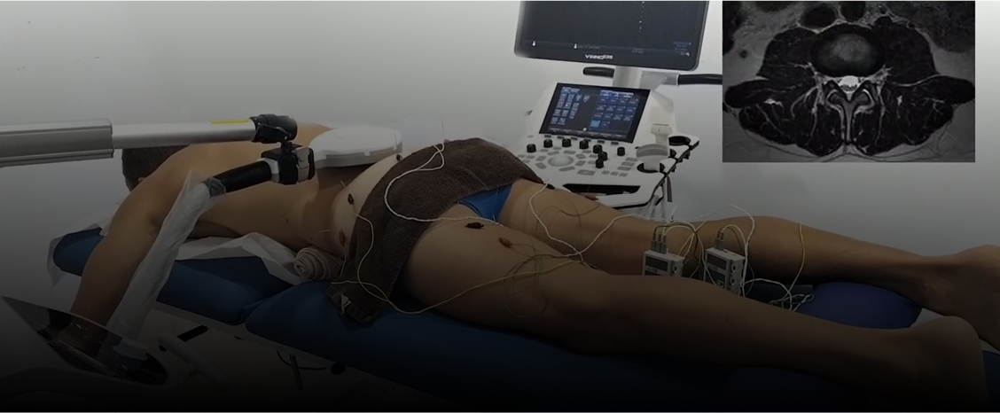
Dynamic ultrasonography examination demonstrating
the full thickness tear and already occurring muscle atrophy
due to misdiagnosis and not referring the patient
to proper diagnostic workup
Demonstration of how very small muscle defect is made and revealed
to be a complete tear with muscle contraction
under diagnostic sonography (not possible with MRI)

Complete tear of rectus femoris
with large hematoma (blood)

Separation of muscle ends due to tear elicited
on dynamic sonography examination


