HPST is developed by Dutch biomedical engineer and a physiotherapist Maarten Prince PT, PHD in collaboration with the Dutch Royal Soccer academy.HSPT has gone rigorous validation process in research scientific community and won several awards.
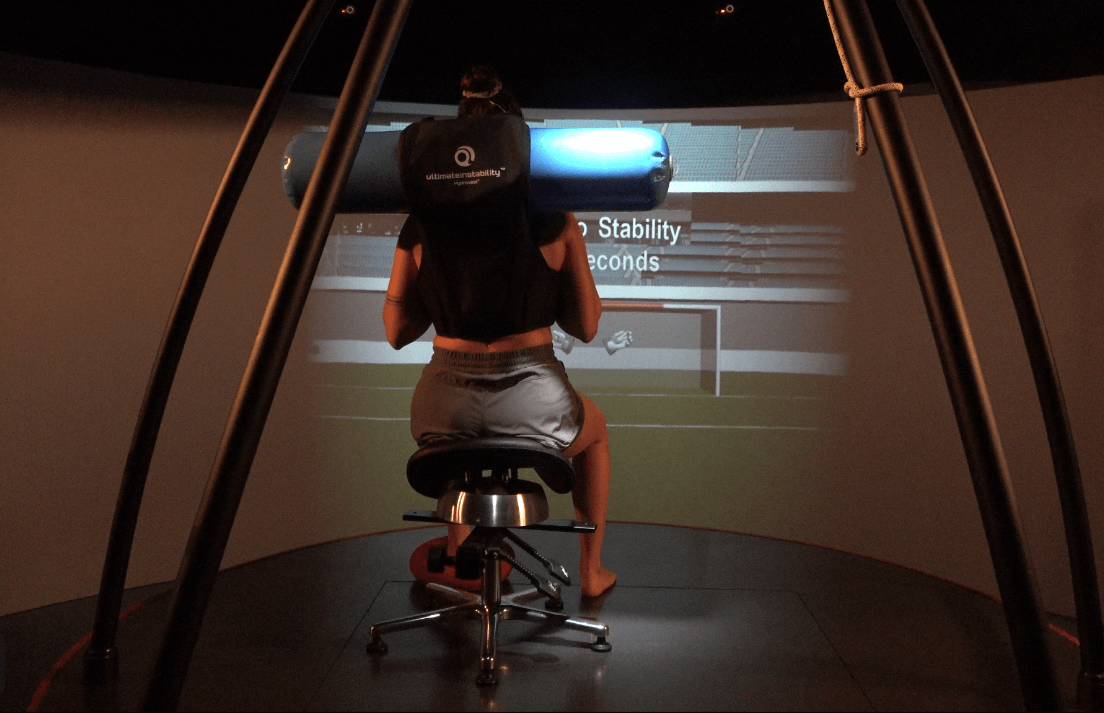
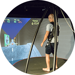
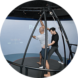
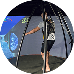
HPST is a perturbation-based test using C.A.R.E.N. (computer assisted rehabilitation environment), with a 6 DoF (six degrees of freedom) instrumented platform, motion capture video and force plate technology. HSPT uses COP (Center of Pressure) velocity and time-to-stability parameters to measure limb stability. This test is used to zero in on ankle and knee stability. It exceeds the capabilities of gait analysis technology for orthopedic and sports medicine disorders, such as ACL and meniscus tears, ankle instability, running deficiencies and other sports-related trauma.
At NYDNRehab we use HSPT as a free standing or in conjunction with gait or running analysis.
Since HSPT is conducted in a matter of minutes we use it to not only for diagnostic purposes but also to establish a base line for monitoring rehabilitation progress.

Below is a prime example of how ultrasound can take the guesswork out of diagnosis.

A bad physical therapy experience is one of the primary causes of unnecessary surgery
In this instance, an athlete was originally diagnosed with minor quadriceps muscle strain and was treated for four weeks, with unsatisfactory results. When he came to our clinic, the muscle was not healing, and the patients’ muscle tissue had already begun to atrophy.
Upon examination using MSUS, we discovered that he had a full muscle thickness tear that had been overlooked by his previous provider. To mitigate damage and promote healing, surgery should have been performed immediately after the injury occurred. Because of misdiagnosis and inappropriate treatment, the patient now has permanent damage that cannot be corrected.
The most important advantage of Ultrasound over MRI imaging is its ability to zero in on the symptomatic region and obtain imaging, with active participation and feedback from the patient. Using dynamic MSUS, we can see what happens when patients contract their muscles, something that cannot be done with MRI. From a diagnostic perspective, this interaction is invaluable.
Dynamic ultrasonography examination demonstrating
the full thickness tear and already occurring muscle atrophy
due to misdiagnosis and not referring the patient
to proper diagnostic workup
Demonstration of how very small muscle defect is made and revealed
to be a complete tear with muscle contraction
under diagnostic sonography (not possible with MRI)

Complete tear of rectus femoris
with large hematoma (blood)

Separation of muscle ends due to tear elicited
on dynamic sonography examination


