
Assessment is fundamental to rehab and training. The initial assessment offers a baseline against which we measure progress. Without concrete baseline measurements, it is nearly impossible to make tangible improvements in performance and function. With Kineo we perform:
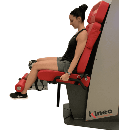
With Kineo, loads can be varied between concentric and eccentric phases, for customized training and rehabilitation programming that gets maximal results.
Eccentric training offers phenomenal strength gains of up to four times those of concentric training. Kineo allows safe and effective eccentric training options to optimize strength and performance.
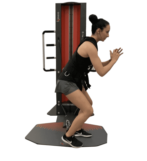
Maximize explosive power and release elastic energy without the impact stress of landing on your back and knees. Rehab lower limb injuries safely and efficiently in Kineo elastic mode. Heal injuries to the Achilles tendon and restore knee joint integrity after ACL reconstruction.
We create customized variable load protocols for functional training, core training, agility drills and more with the Kineo variable resistance cable-pulley system. Kineo lets us design a variable load curve based on the individual needs of each patient. Kineo allows us to:
Kineo’s viscous adaptive load method allows for functional movement without exceeding the pain threshold. Gain the benefits of water training without getting wet!
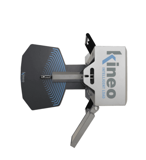
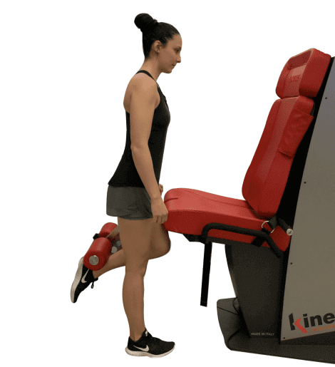
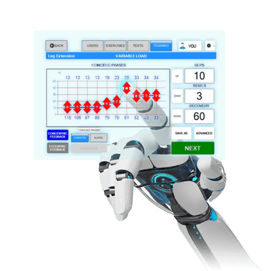
Get visual and auditory feedback as you exercise, to keep you motivated and help you reach your goals.
Dr. Kalika is currently a certified member of:
American Institute of Ultrasound Medicine

Active member of ISMST
International Society of Extra Corporeal Shockwave Therapy
Active member of GCMAS
Gait and Clinical Movement Analysis Society
Active member of NASS
North American Spine Society
Active member of IADMS
International Association of Dance Medicine and Science
Active member of Virtual Rehabilitation Society
Active member of ASRA
American Society of Regional Anesthesia and Pain Medicine
American Academy
Association of Orthopedic Medicine

Active member of Interventional Orthobiologics Foundation


Verified Expert Profiles
Dr. Lev Kalika is a world-recognized expert in musculoskeletal medicine. with 20+ years of clinical experience in diagnostic musculoskeletal ultrasonography, rehabilitative sports medicine and conservative orthopedics. In addition to operating his clinical practice in Manhattan, he regularly publishes peer-reviewed research on ultrasound-guided therapies and procedures. He serves as a peer reviewer for Springer Nature.
Dr. Kalika is an esteemed member of multiple professional organizations, including:
Below is a prime example of how ultrasound can take the guesswork out of diagnosis.

A bad physical therapy experience is one of the primary causes of unnecessary surgery
In this instance, an athlete was originally diagnosed with minor quadriceps muscle strain and was treated for four weeks, with unsatisfactory results. When he came to our clinic, the muscle was not healing, and the patients’ muscle tissue had already begun to atrophy.
Upon examination using MSUS, we discovered that he had a full muscle thickness tear that had been overlooked by his previous provider. To mitigate damage and promote healing, surgery should have been performed immediately after the injury occurred. Because of misdiagnosis and inappropriate treatment, the patient now has permanent damage that cannot be corrected.
The most important advantage of Ultrasound over MRI imaging is its ability to zero in on the symptomatic region and obtain imaging, with active participation and feedback from the patient. Using dynamic MSUS, we can see what happens when patients contract their muscles, something that cannot be done with MRI. From a diagnostic perspective, this interaction is invaluable.
Dynamic ultrasonography examination demonstrating
the full thickness tear and already occurring muscle atrophy
due to misdiagnosis and not referring the patient
to proper diagnostic workup
Demonstration of how very small muscle defect is made and revealed
to be a complete tear with muscle contraction
under diagnostic sonography (not possible with MRI)

Complete tear of rectus femoris
with large hematoma (blood)

Separation of muscle ends due to tear elicited
on dynamic sonography examination


