Three years. It’s what it took for a 28-year-old competitive runner to suffer from lateral calf pain and not be able to train at his full capacity. He’d had the MRI. He had already been to several doctors. Each time the imaging was fine, and he was informed that there was no structural problem. Yet the pain recurred whenever he ran.
A repeat MRI or a different radiologist did not ultimately pinpoint the problem. Dynamic musculoskeletal ultrasound, using real-time imaging during the patient’s movements, was the test that revealed a peroneal nerve gliding disorder that had been missed by all previous tests.
| Patient | 28-year-old male competitive runner |
|---|---|
| Chief complaint | Lateral calf pain, worse with running |
| Duration | 3 years |
| Prior testing | MRI (reported normal), clinical evaluations |
| Key finding | Peroneal nerve snapping and restricted glide at the proximal fibular tunnel on dynamic ultrasound |
| Treatment | Ultrasound-guided hydrodissection at the fibular tunnel |
| Outcome | Symptom-free at 10-week follow-up; returned to full running volume |
The athlete experiences lateral calf pain during longer training sessions. The pattern remained the same over the three years: pain would set in on running, but would abate when rested, and would return when mileage was increased. It was not a single event that was traumatic. No ankle sprain. No obvious mechanism.
He had already undergone the usual work-up. An MRI of the lower leg revealed no stress fracture, muscle tear or tendon pathology. No clinical findings were suggestive of structural lesion. There was no diagnosis and treatment remained general: rest, stretching, and gradual return to training, which continued to fail.
Our clinic’s first evaluation consisted of neural tension testing and regional palpation. He complained of the same symptoms on palpation of the proximal fibular tunnel, which suggested that this was a problem with the peroneal nerve. That discovery led to the decision to image dynamically, instead of repeating the static workup that had come up blank.
MRI is a great instrument for identifying structural lesions like muscle tears, bone stress, tendon degeneration. It’s not that it doesn’t do well at capturing the behavior of tissues under load, or during movement. The images are acquired with the patient lying still.
This is especially true of the peroneal nerve at the fibular head. The nerve can be in a normal-appearing position at rest in its tunnel. This issue only becomes apparent when the muscles around the tunnel are contracting and the architecture of the tunnel changes.
This patient’s problem was due to the soleus-peroneus longus fibrous arch complex (fibrous band which is the roof of the fibular tunnel). In motion, it may change and compress the nerve. When the patient is still, the MRI shows nothing abnormal. The nerve is there. The arch is there. No compression is visible because no compression is taking place.
Figure 1: Ultrasound comparison – normal fibular tunnel architecture versus symptomatic nerve position at rest and under load
The MRI did not provide any information that ultrasound could: the peroneal nerve was snapping at the proximal fibular tunnel while the ankle and foot was moving.
The nerve was abnormally displaced with every movement of plantarflexion and dorsiflexion of the ankle. It did not slide easily down its sheath but caught and released with each step, a mechanical process. This is known as a peroneal nerve glide disorder. Patients frequently complain of a snap, click or catch at the outside of the knee or at the top of the calf, and the pain is likely to gradually get worse over a run.
Figure 2: Dynamic ultrasound sequence demonstrating peroneal nerve displacement and snapping at the fibular tunnel during active plantarflexion-dorsiflexion.
Changes consistent with chronic mechanical stress were also seen in the nerve’s fascicular architecture. Nerve fibers are grouped together into fascicles within a nerve. In cases of repeated compression or torsion of a nerve, the internal fascicular pattern can become disrupted and when the pattern was seen under high resolution ultrasound, this was not an incidental anatomical variant but an active clinically relevant problem.
Underlying mechanics: the concept of muscular-aponeurotic encroachment. During running, the two muscles, the peroneus longus and the soleus, pull on the nerve through their fibrous arch, which is like a closing gate. The discomfort is created over time, with each step, and is the reason why it hurts more during a run and feels better when you rest.
This discovery altered the clinical picture altogether. There was now a clear, well-defined, mechanical problem to be fixed, not a lateral calf complaint with no mechanical target.
A procedure selected was ultrasound guided hydrodissection at the fibular tunnel. Hydrodissection is a technique in which a small amount of fluid (usually saline solution) is injected around a nerve under ultrasound guidance. The fluid will separate the nerve from the surrounding tissues that are pulling or squeezing the nerve, allowing it to move freely.
The clinician guided the needle by live ultrasound imaging to the exact location of nerve entrapment. The fluid was given to reach the nerve and the fibrous arch complex to break the adhesion. Immediate reevaluation of nerve movement was done after the procedure.
Figure 3: Post-hydrodissection ultrasound showing restored peroneal nerve glide through the fibular tunnel
The patient was symptom free and back to his full training volume at 10-week follow up. A single targeted procedure yielded long-lasting pain relief in that time, despite the fact that three years of running had been interrupted and a diagnostic procedure had been performed with no success.
The change was statistically significant. He said there was no pain in his lateral calf when running, no snapping in the calf, and no changes to the intensity or duration of his training. That’s a considerable functional recovery, not just a decrease in pain scores, for a competitive runner who had been limited for three years.
It is unclear whether the hydrodissection was sufficient to remove the underlying biomechanical tendency to nerve compression or whether it was sufficient to provide enough mechanical relief for normal neuromechanical adaptation to occur over the weeks following the procedure. A clinical signal of 10 weeks is good. It is not a permanent verdict.
Yes, and this is an example of a case. MRI images show the anatomy in its normal state. Peroneal nerve entrapment at the fibular tunnel is frequently a dynamic problem, the nerve becoming compressed only during movement when the muscles around the fibular tunnel contract and compress the nerve. MRI is not helpful in excluding a nerve glide disorder.
Dynamic ultrasound is different from standard ultrasound because the patient moves the affected joint or limb while the ultrasound is performed. Clinicians are able to observe tendons, nerves and surrounding tissues as they function during movement. In nerve gliding problems, this is sometimes the only imaging technique that reveals the pathology.
In hydrodissection, a clinician guides a needle using real-time ultrasound to a specific area around a nerve, and injects a small amount of fluid into the area to physically separate the nerve from scar tissue or fibrous tissue that is limiting its movement. It is used when a nerve is not just inflamed, but is also “tethered”.
Yes. Peroneal nerve irritation at the fibular tunnel is a very specific cause of lateral calf pain in runners that is underdiagnosed, and that will improve with rest and recur with continued running despite normal imaging. If you have this pattern for months and your MRI is not remarkable, then you should consider a nerve cause.
Most often, it is due to soleus strain, fibular stress fracture, lateral compartment syndrome, and peroneal tendinopathy. Peroneal nerve entrapment at the proximal fibular tunnel is a less commonly known condition that causes a characteristic pain in the lateral calf, which worsens with activity. During running the nerve may be compressed intermittently by the fibrous arch created by the soleus and peroneus longus, especially at high speeds and distances.
Normal MRI does not equal normal diagnosis.
Dr. Lev Kalika is a world-recognized expert in musculoskeletal medicine. with 20+ years of clinical experience in diagnostic musculoskeletal ultrasonography, rehabilitative sports medicine and conservative orthopedics. In addition to operating his clinical practice in Manhattan, he regularly publishes peer-reviewed research on ultrasound-guided therapies and procedures. He serves as a peer reviewer for Springer Nature.
Dr. Kalika is an esteemed member of multiple professional organizations, including:

Below is a prime example of how ultrasound can take the guesswork out of diagnosis.

A bad physical therapy experience is one of the primary causes of unnecessary surgery

In this instance, an athlete was originally diagnosed with minor quadriceps muscle strain and was treated for four weeks, with unsatisfactory results. When he came to our clinic, the muscle was not healing, and the patients’ muscle tissue had already begun to atrophy.
Upon examination using MSUS, we discovered that he had a full muscle thickness tear that had been overlooked by his previous provider. To mitigate damage and promote healing, surgery should have been performed immediately after the injury occurred. Because of misdiagnosis and inappropriate treatment, the patient now has permanent damage that cannot be corrected.
The most important advantage of Ultrasound over MRI imaging is its ability to zero in on the symptomatic region and obtain imaging, with active participation and feedback from the patient. Using dynamic MSUS, we can see what happens when patients contract their muscles, something that cannot be done with MRI. From a diagnostic perspective, this interaction is invaluable.
Dynamic ultrasonography examination demonstrating
the full thickness tear and already occurring muscle atrophy
due to misdiagnosis and not referring the patient
to proper diagnostic workup
Demonstration of how very small muscle defect is made and revealed
to be a complete tear with muscle contraction
under diagnostic sonography (not possible with MRI)
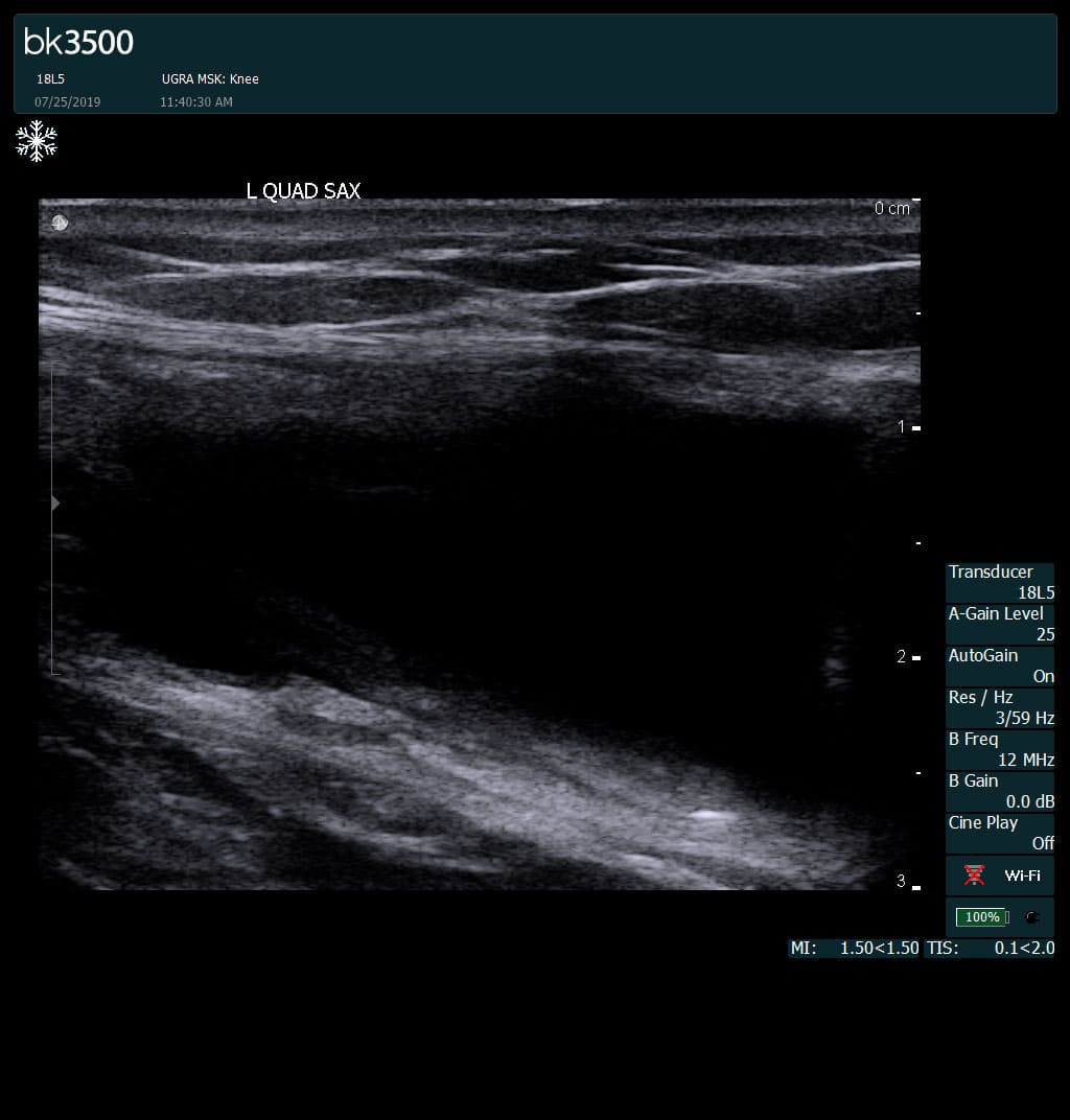
Complete tear of rectus femoris
with large hematoma (blood)
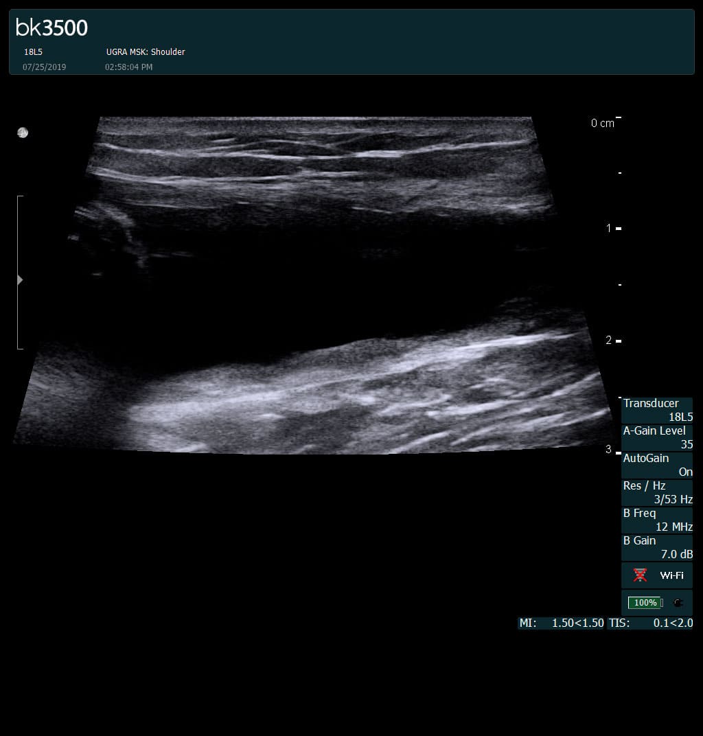
Separation of muscle ends due to tear elicited
on dynamic sonography examination

 Request Telehealth