Our patient is a 35 year-old female complaining of persistent pain and tingling over the right chest area. The pain intensified during loading exercises like bench presses, and during pushing movements.
The patient had seen two orthopedists who were unable to explain her symptoms. MR imaging came back negative. EMG/MCV evaluations performed by two separate neurologists were also negative. The patient was sent to a physical therapist, but the therapy only aggravated her pain. She came to us for help.
Clinical Exam
During the clinical exam, we noticed a scar over the right clavicle. The patient informed us that she had undergone open reduction and internal fixation (ORIF) surgery in 2013 to repair a right clavicle fracture sustained while skiing. The procedure included the installation of a metal plate and screws to secure the clavicle.
collapsed fascial planes adherent to clavicular plate; enlarged/thickened right supraclavicular nerve branches in scar tissue.
Our differential diagnosis indicated that her pain could only come from three sources:
Considering that her scar was directly over the right clavicle, it seemed obvious to look into the supraclavicular nerves.
Ultrasound Exam
An examination using high-resolution diagnostic ultrasound revealed thickening of the anterior trapezius fascial planes, with one surascapular branch tethered against the sharp edge of the plate. Nerve branches were enlarged and embedded in scar tissue. We concluded that the patient had developed scapular dyskinesis and fascial thickening over time, eventually leading to supraclavicular nerve entrapment.

We performed ultrasound-guided hydrodissection of the right supraclavicular nerve branches and surrounding fibrotic fascia using a 5 % dextrose solution, targeting the fascial plate interface and areas adherent to the metal hardware. It took only a single hydrodissection procedure to produce 100% symptom relief.
In this case, doctors were perplexed and unable to pinpoint the underlying source of the patient’s pain. Considering the presence of fascial thickening and scar tissue, physical therapy exercises meant to strengthen the upper body only increased pressure on the affected nerves. The doctors completely overlooked the patient’s previous surgery as a potential contributor to her current symptoms.
It is not uncommon for the body to form scar tissue after a fracture and surgery. In this case, the patient’s condition gradually worsened over time, intensifying pressure on her subclavicular nerves. We immediately suspected that her symptoms were related to her old injury and surgery, prompting us to examine the subclavicular space. It only took a quick and minimally invasive ultrasound-guided procedure to completely eliminate her symptoms.
Dr. Lev Kalika is a world-recognized expert in musculoskeletal medicine. with 20+ years of clinical experience in diagnostic musculoskeletal ultrasonography, rehabilitative sports medicine and conservative orthopedics. In addition to operating his clinical practice in Manhattan, he regularly publishes peer-reviewed research on ultrasound-guided therapies and procedures. He serves as a peer reviewer for Springer Nature.
Dr. Kalika is an esteemed member of multiple professional organizations, including:

Below is a prime example of how ultrasound can take the guesswork out of diagnosis.

A bad physical therapy experience is one of the primary causes of unnecessary surgery

In this instance, an athlete was originally diagnosed with minor quadriceps muscle strain and was treated for four weeks, with unsatisfactory results. When he came to our clinic, the muscle was not healing, and the patients’ muscle tissue had already begun to atrophy.
Upon examination using MSUS, we discovered that he had a full muscle thickness tear that had been overlooked by his previous provider. To mitigate damage and promote healing, surgery should have been performed immediately after the injury occurred. Because of misdiagnosis and inappropriate treatment, the patient now has permanent damage that cannot be corrected.
The most important advantage of Ultrasound over MRI imaging is its ability to zero in on the symptomatic region and obtain imaging, with active participation and feedback from the patient. Using dynamic MSUS, we can see what happens when patients contract their muscles, something that cannot be done with MRI. From a diagnostic perspective, this interaction is invaluable.
Dynamic ultrasonography examination demonstrating
the full thickness tear and already occurring muscle atrophy
due to misdiagnosis and not referring the patient
to proper diagnostic workup
Demonstration of how very small muscle defect is made and revealed
to be a complete tear with muscle contraction
under diagnostic sonography (not possible with MRI)
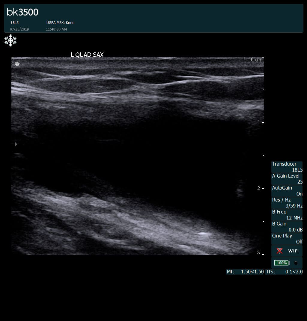
Complete tear of rectus femoris
with large hematoma (blood)
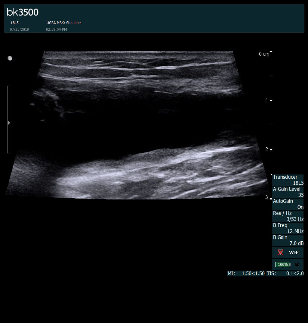
Separation of muscle ends due to tear elicited
on dynamic sonography examination


 Request Telehealth