Our patient is a 79 year-old female whose son escorted her to our clinic. She was only able to lift her arm to about 30° before losing strength, and deep shoulder pain was shooting down to mid-arm.
Eight months earlier the patient had been diagnosed with “frozen shoulder” (aka adhesive capsulitis) by an orthopedist who made the diagnosis based on symptoms alone, with no exam or imaging.
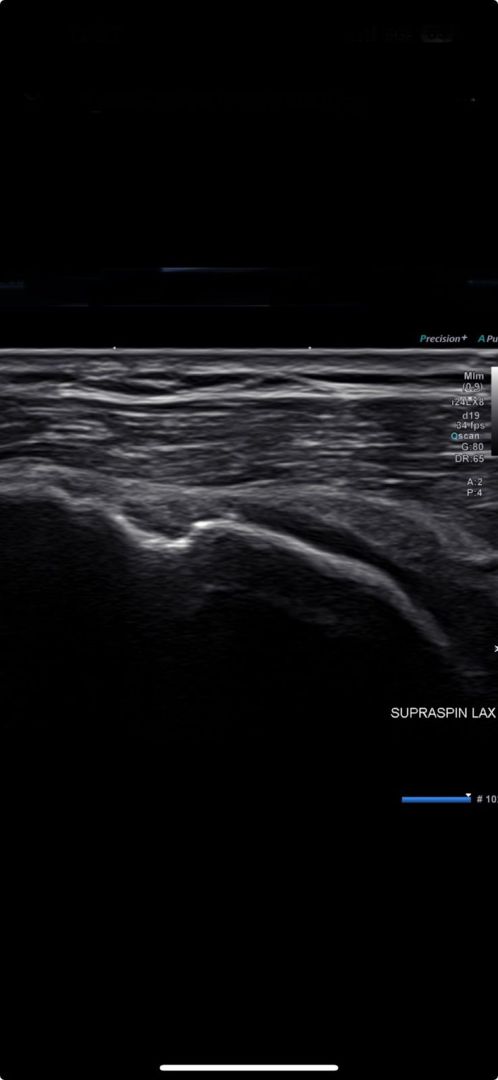
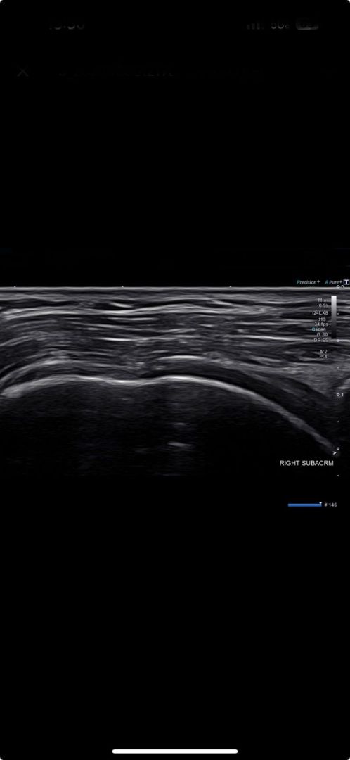
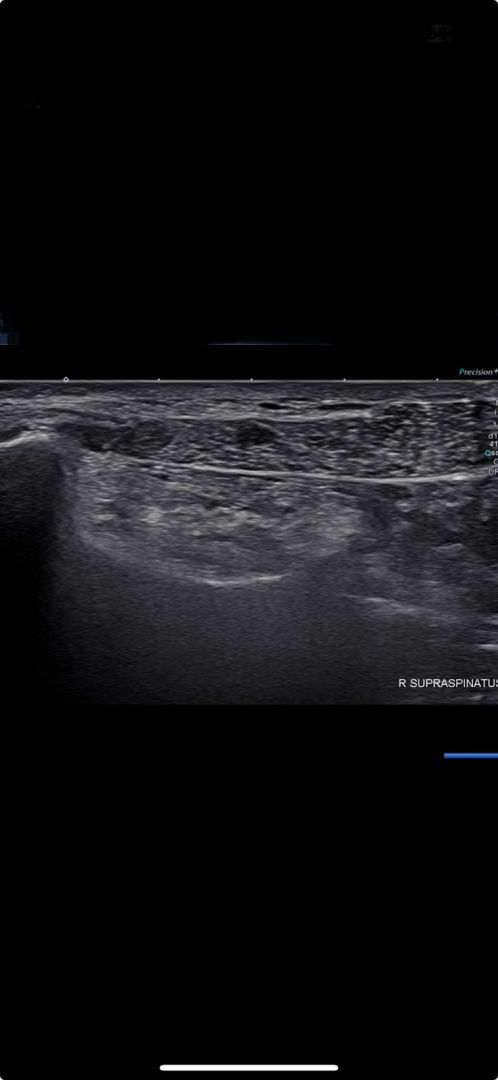
After reviewing her health history and conducting a physical exam, we used high-resolution diagnostic ultrasonography to visualize the patient’s arm and shoulder region.
Reduced shoulder range of motion does not always indicate adhesive capsulitis, and
structural causes must be ruled out. The delay in accurate diagnosis prolonged the patient’s suffering for eight months, and the condition no doubt worsened along the way. The delay could have been prevented if the original orthopedist had taken the time to conduct a hands-on physical exam and ordered imaging.

Verified Expert Profiles
Dr. Lev Kalika is a world-recognized expert in musculoskeletal medicine. with 20+ years of clinical experience in diagnostic musculoskeletal ultrasonography, rehabilitative sports medicine and conservative orthopedics. In addition to operating his clinical practice in Manhattan, he regularly publishes peer-reviewed research on ultrasound-guided therapies and procedures. He serves as a peer reviewer for Springer Nature.
Dr. Kalika is an esteemed member of multiple professional organizations, including:
Below is a prime example of how ultrasound can take the guesswork out of diagnosis.

A bad physical therapy experience is one of the primary causes of unnecessary surgery
In this instance, an athlete was originally diagnosed with minor quadriceps muscle strain and was treated for four weeks, with unsatisfactory results. When he came to our clinic, the muscle was not healing, and the patients’ muscle tissue had already begun to atrophy.
Upon examination using MSUS, we discovered that he had a full muscle thickness tear that had been overlooked by his previous provider. To mitigate damage and promote healing, surgery should have been performed immediately after the injury occurred. Because of misdiagnosis and inappropriate treatment, the patient now has permanent damage that cannot be corrected.
The most important advantage of Ultrasound over MRI imaging is its ability to zero in on the symptomatic region and obtain imaging, with active participation and feedback from the patient. Using dynamic MSUS, we can see what happens when patients contract their muscles, something that cannot be done with MRI. From a diagnostic perspective, this interaction is invaluable.
Dynamic ultrasonography examination demonstrating
the full thickness tear and already occurring muscle atrophy
due to misdiagnosis and not referring the patient
to proper diagnostic workup
Demonstration of how very small muscle defect is made and revealed
to be a complete tear with muscle contraction
under diagnostic sonography (not possible with MRI)

Complete tear of rectus femoris
with large hematoma (blood)

Separation of muscle ends due to tear elicited
on dynamic sonography examination


