Our patient is a young adult female athlete who was undergoing rehabilitation after reconstructive knee surgery. The patient came to us complaining of post-surgery knee instability.
The patient’s surgery had been performed by a top knee surgeon at a prestigious Qatari sports medicine hospital – a key destination for injured elite athletes from around the world. After a successful procedure, she had begun rehabilitation with some of the world’s top sports physiotherapists. Post-surgical MRI showed normal MCL and LCL ligaments. However, the patient reported that the knee did not feel stable, and she came to us for help.
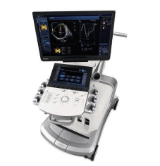
After a standard clinical exam, we conducted dynamic imaging of the knee using high-resolution ultrasound, along with gait analysis and stress testing for knee stability. Both the MCL and the LCL appeared to be intact, but dynamic imaging revealed a loss of tension in the ligaments. We concluded that the patient had mild Ehlers-Danlos syndrome, causing hypermobile joints that needed specialized treatment.
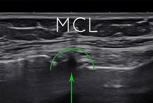
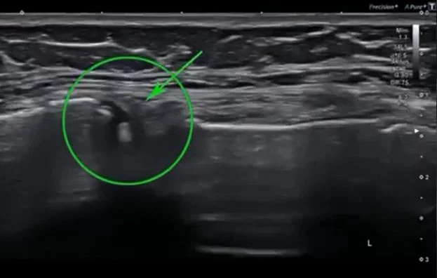
We treated the patient’s ligaments with a series of Prolotherapy injections, along with 3 months of EDS-specific physical rehabilitation. She reported significant improvement in knee stability, and results from a 3D gait analysis showed measurable improvements in multiple kinematic and spatiotemporal parameters.
Once we improved her knee stability, the patient returned to the Doha clinic to complete her rehabilitation protocol, with our recommendation that the rehab be conducted by a specialist experienced in working with hypermobile and EDS patients.
When treating patients with joint hypermobility, the joint capsule often appears normal on static imaging like MRI. However, when observing the joint in motion with dynamic ultrasound imaging, we were able to detect a loss of tension in the knee’s supporting structures, affecting overall stability. Such cases require specialized treatment by a clinician with expertise and experience working with hypermobile patients.
For many athletes, knee surgery is performed as soon as possible after a knee injury to expedite their return to sport. But in our opinion, patients with EDS should avoid joint surgery unless it is absolutely necessary, since surgical procedures can exacerbate joint instability and increase the risk of further injury.
Dr. Lev Kalika is a world-recognized expert in musculoskeletal medicine. with 20+ years of clinical experience in diagnostic musculoskeletal ultrasonography, rehabilitative sports medicine and conservative orthopedics. In addition to operating his clinical practice in Manhattan, he regularly publishes peer-reviewed research on ultrasound-guided therapies and procedures. He serves as a peer reviewer for Springer Nature.
Dr. Kalika is an esteemed member of multiple professional organizations, including:

Below is a prime example of how ultrasound can take the guesswork out of diagnosis.

A bad physical therapy experience is one of the primary causes of unnecessary surgery

In this instance, an athlete was originally diagnosed with minor quadriceps muscle strain and was treated for four weeks, with unsatisfactory results. When he came to our clinic, the muscle was not healing, and the patients’ muscle tissue had already begun to atrophy.
Upon examination using MSUS, we discovered that he had a full muscle thickness tear that had been overlooked by his previous provider. To mitigate damage and promote healing, surgery should have been performed immediately after the injury occurred. Because of misdiagnosis and inappropriate treatment, the patient now has permanent damage that cannot be corrected.
The most important advantage of Ultrasound over MRI imaging is its ability to zero in on the symptomatic region and obtain imaging, with active participation and feedback from the patient. Using dynamic MSUS, we can see what happens when patients contract their muscles, something that cannot be done with MRI. From a diagnostic perspective, this interaction is invaluable.
Dynamic ultrasonography examination demonstrating
the full thickness tear and already occurring muscle atrophy
due to misdiagnosis and not referring the patient
to proper diagnostic workup
Demonstration of how very small muscle defect is made and revealed
to be a complete tear with muscle contraction
under diagnostic sonography (not possible with MRI)
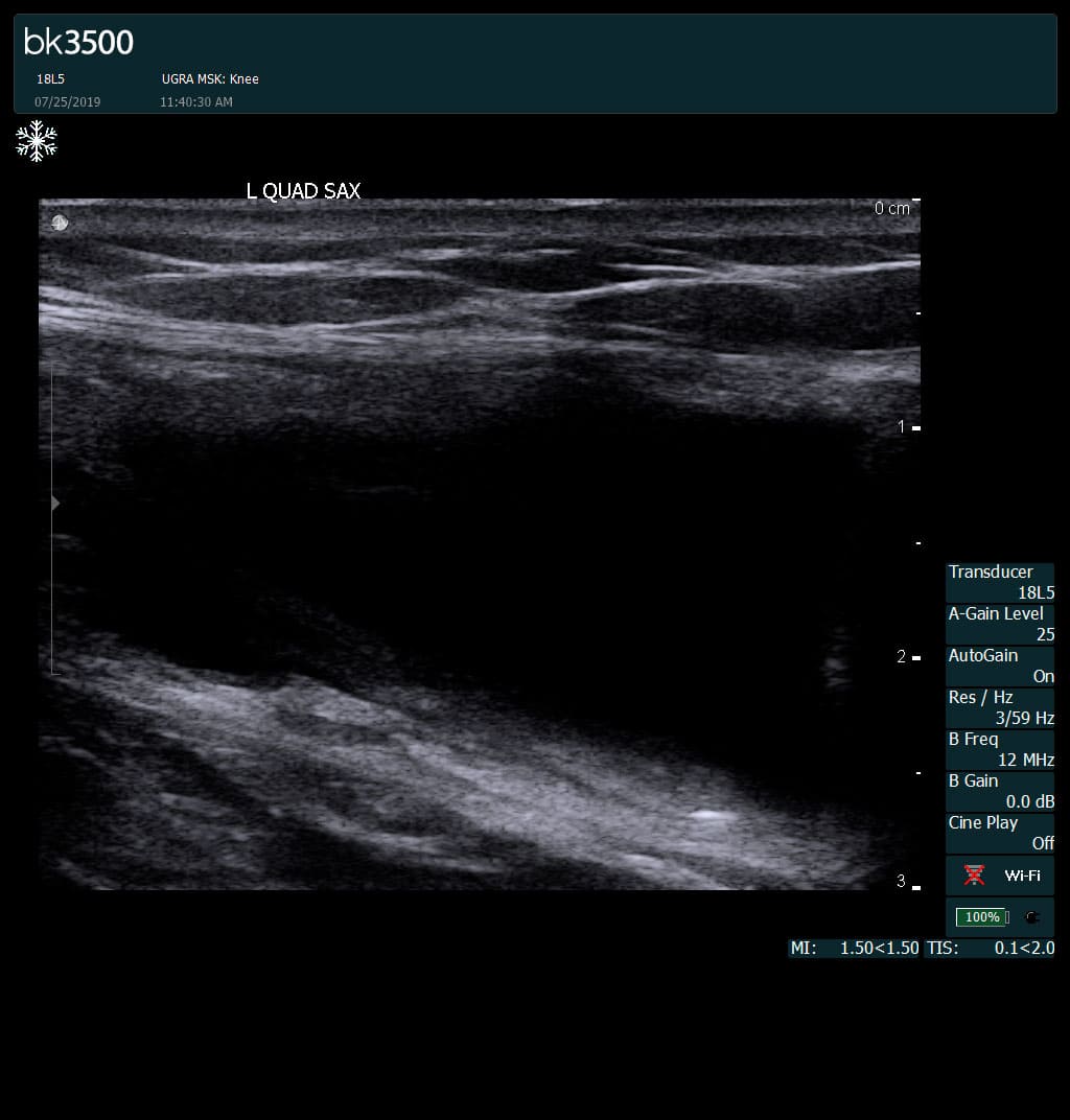
Complete tear of rectus femoris
with large hematoma (blood)
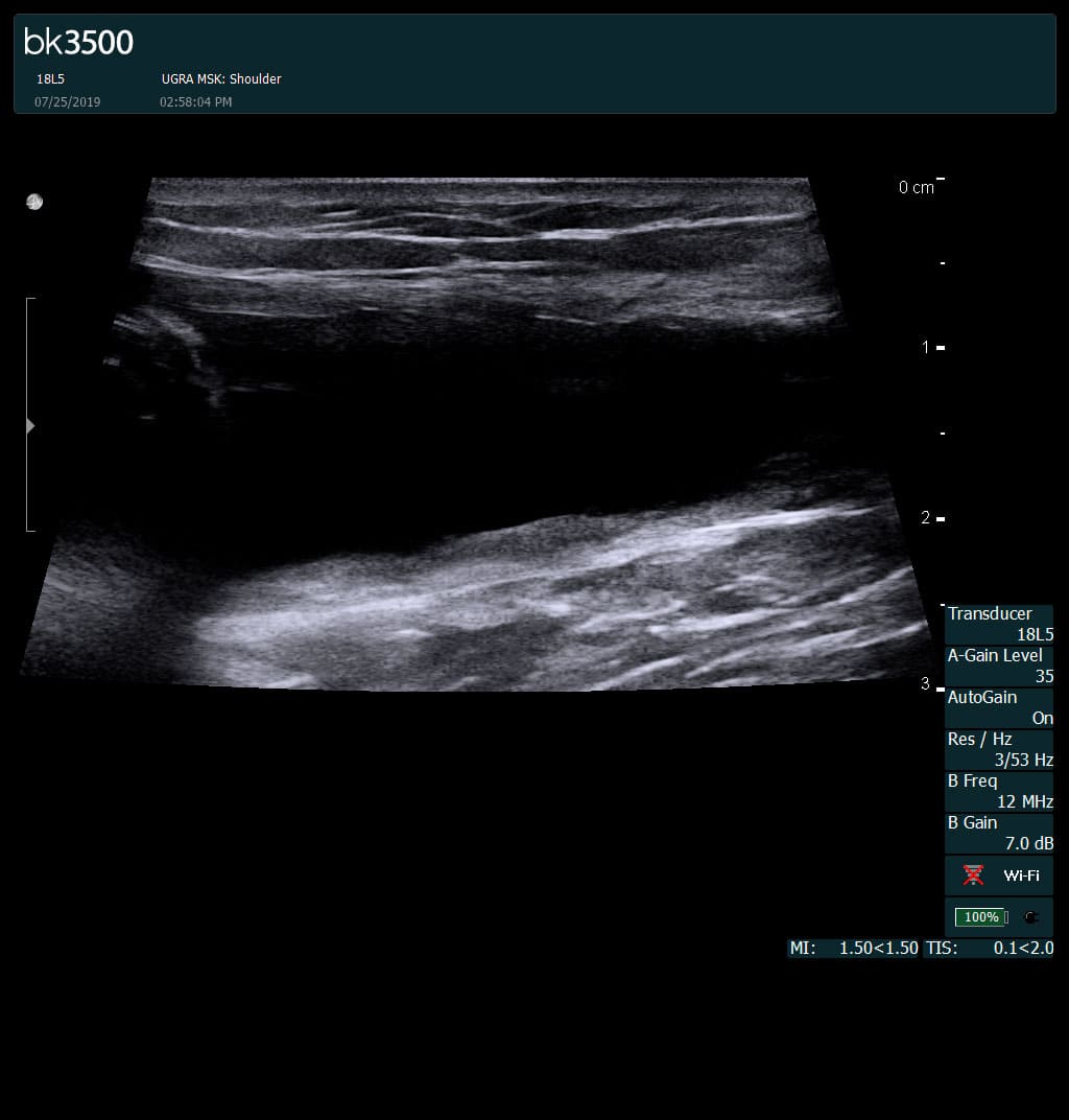
Separation of muscle ends due to tear elicited
on dynamic sonography examination


 Request Telehealth