Our patient is a 26 year-old female and former ballet dancer, complaining of hip, groin, and lower back pain. She presented with multiple symptoms of Ehlers-Danlos syndrome and had recently undergone surgery for endometriosis.
The patient had previously seen multiple physical therapists, with no satisfactory results. She had received bilateral platelet rich plasma (PRP) injections into both of her adductor longus insertion sites, but continued to experience groin pain. MRI had revealed multiple bulging discs, which were treated with epidural steroid injections,
with no positive results.
Clinical Exam
Our clinical exam included a medical history review and physical exam, followed by standardized testing.
Our most remarkable findings were:
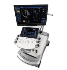
Imaging with high resolution ultrasound revealed a lesion of the Pyramidalis-Anterior Pubic Ligament-Adductor Longus Complex (PLAC), a common athletic injury where the proximal adductor tendon tears away from the pubic bone. The pubic symphysis, suprapubic ligaments, and SI joint ligaments appeared normal. We also observed multiple densifications along the abdominal sheath.
The patient was unable to engage her abdominal muscles adequately to stabilize her pelvis, and her hypermobile ligaments could not stabilize her anterior or posterior pelvic ring. Despite normal appearance on ultrasound, the SI ligaments were not able to compensate for lack of abdominal engagement due to multiple abdominal incisions from her previous endometriosis surgery.
It soon became clear that her hypermobile hips were placing stress on the SI ligaments, and that her symptoms of groin pain were likely related to SI joint dysfunction. In this case, the SI ligaments were pain generators, not only for lower back pain, but also for groin pain.
The patient’s SI ligaments and pubic symphysis were injected with a Prolotherapy solution of D5W, which reduced all of her pain symptoms by 70%, making physical therapy possible. We also used fascial manipulation to restore gliding of the recto-abdominal sheath, which restored coordinated abdominal muscle activation for improved stabilization.
When evaluating patients with lumbopelvic pain, we need to consider factors affecting pelvic stability, even when imaging of ligaments appears normal. This is especially in cases of joint hypermobility or EDS. In such cases, imaging alone is insufficient – the clinical evaluation and stress testing results must be relied on to guide professional reasoning.
Dr. Lev Kalika is a world-recognized expert in musculoskeletal medicine. with 20+ years of clinical experience in diagnostic musculoskeletal ultrasonography, rehabilitative sports medicine and conservative orthopedics. In addition to operating his clinical practice in Manhattan, he regularly publishes peer-reviewed research on ultrasound-guided therapies and procedures. He serves as a peer reviewer for Springer Nature.
Dr. Kalika is an esteemed member of multiple professional organizations, including:

Below is a prime example of how ultrasound can take the guesswork out of diagnosis.

A bad physical therapy experience is one of the primary causes of unnecessary surgery

In this instance, an athlete was originally diagnosed with minor quadriceps muscle strain and was treated for four weeks, with unsatisfactory results. When he came to our clinic, the muscle was not healing, and the patients’ muscle tissue had already begun to atrophy.
Upon examination using MSUS, we discovered that he had a full muscle thickness tear that had been overlooked by his previous provider. To mitigate damage and promote healing, surgery should have been performed immediately after the injury occurred. Because of misdiagnosis and inappropriate treatment, the patient now has permanent damage that cannot be corrected.
The most important advantage of Ultrasound over MRI imaging is its ability to zero in on the symptomatic region and obtain imaging, with active participation and feedback from the patient. Using dynamic MSUS, we can see what happens when patients contract their muscles, something that cannot be done with MRI. From a diagnostic perspective, this interaction is invaluable.
Dynamic ultrasonography examination demonstrating
the full thickness tear and already occurring muscle atrophy
due to misdiagnosis and not referring the patient
to proper diagnostic workup
Demonstration of how very small muscle defect is made and revealed
to be a complete tear with muscle contraction
under diagnostic sonography (not possible with MRI)
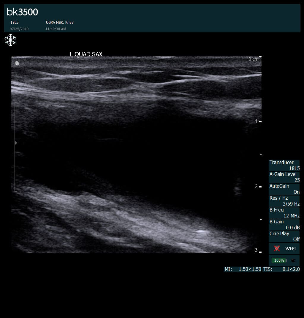
Complete tear of rectus femoris
with large hematoma (blood)
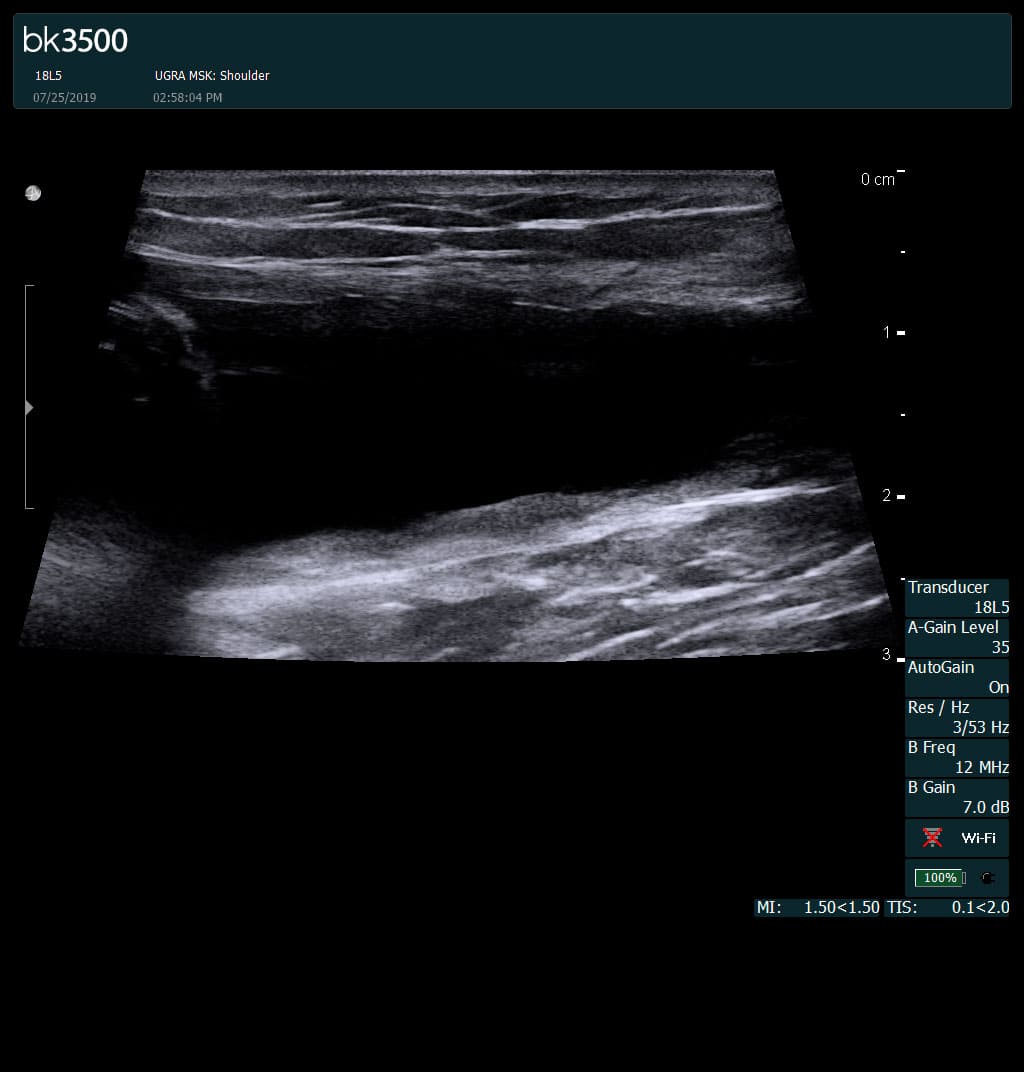
Separation of muscle ends due to tear elicited
on dynamic sonography examination


 Request Telehealth