Our patient is a 65 year-old male complaining of chronic low back pain located above the pelvic rim. He complained of stiffness, with difficulty walking and bending, although he reported no pain with sitting.
The patient brought with him an MRI that indicated multilevel severe disc disease with spinal stenosis. He had seen multiple doctors and physical therapists, had years of physical therapy, tried acupuncture, and had multiple epidural steroid injections, none of which resolved his chronic low back pain. He had consulted with three different surgeons, all of whom wanted to operate on his spine.
During the clinical exam it became clear that none of the patient’s symptoms matched disc or stenosis pain. There was tenderness on palpation of the regions of the spine where he complained of pain, prompting us to take a closer look.
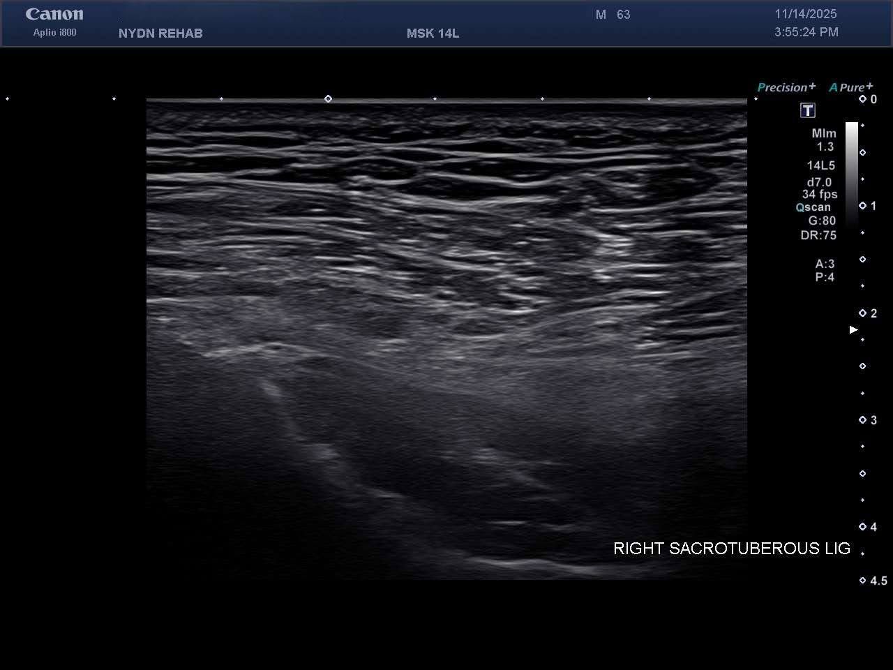
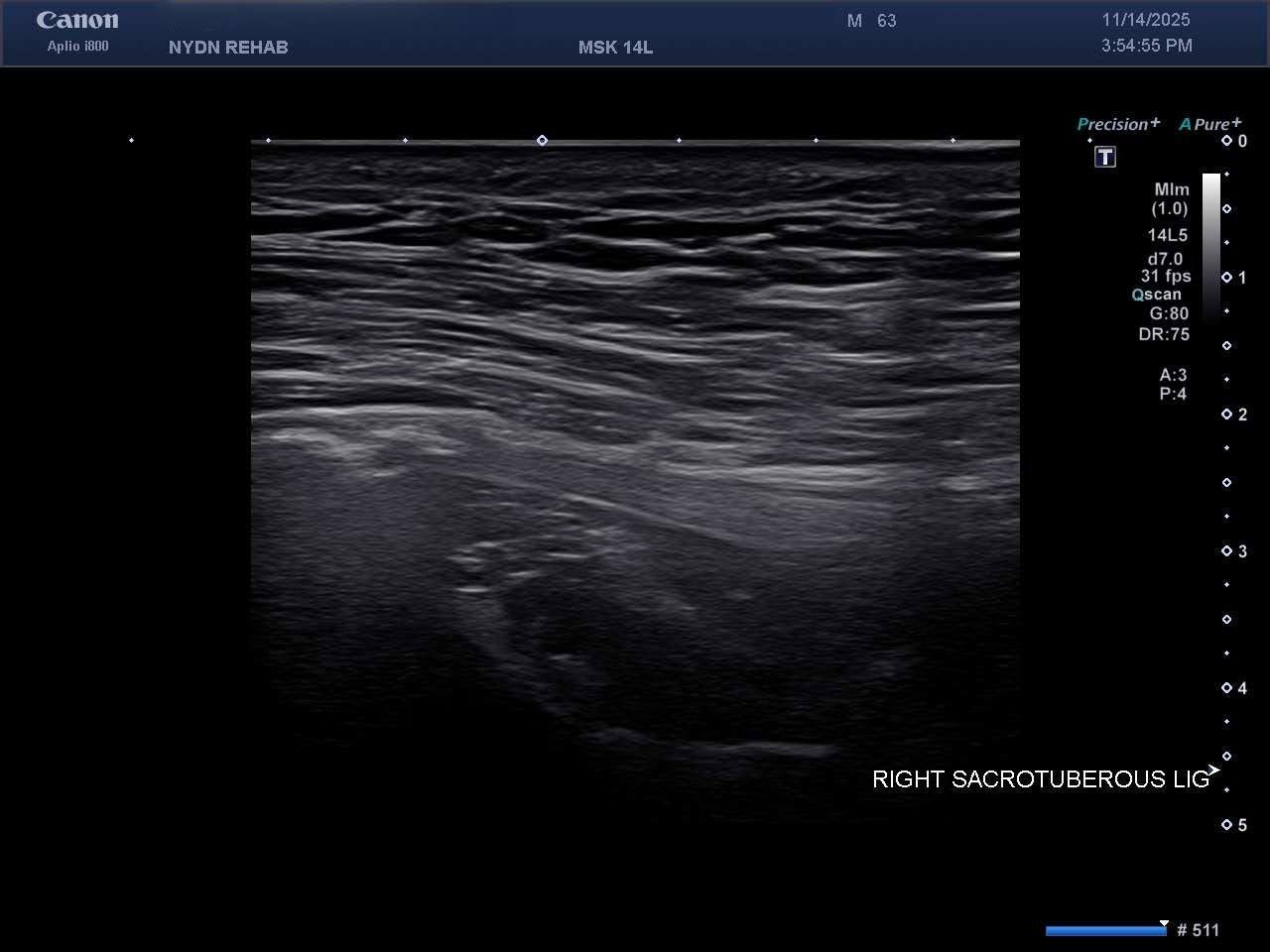
Using high resolution ultrasound imaging, we found:
From our findings we determined that his pain was not discogenic – it was caused by soft tissues and mechanical overload.
Ultrasound Images of SIJ Ligaments

We initially used ultrasound guided focal shockwave therapy, along with extracorporeal mechanotransduction therapy (EMTT) targeting the enthesopathic sacroiliac joint and its iliocostal attachment. In just 3 weekly sessions, the patient’s mobility improved by 80%.
After weeks 4 and 5, the pain was completely gone, and he was now able to walk for an hour at a time. However, surprising new symptoms emerged just above his sit bones and behind his hips.
These findings indicated a failure of tensegrity in the SI joint, which made sense, since the multifidi spinal muscles were atrophied and tension had been lost in the lumbodorsal fascia, increasing stress on the sacroiliac ligaments and disrupting the posterior chain. This all pointed to the real problem: pelvic ligament and posterior-lateral hip tendon/capsule failure.
Once we identified the problem, we delivered one treatment of Prolotherapy followed by PRP injections into the pelvic ligaments, hip tendons, joint capsule, and multifidi. We also trained the patient in SI joint force closure, multifidi activation, intra-abdominal pressure activation of the core, and lateral hip strengthening.
Our treatment approach fully restored stability and tensegrity, and completely eliminated the patient’s low back, hip and pelvic pain – all without surgery. The patient was able to return to his home in the Catskills of Upstate New York, where he walks for miles every day without pain.
Misdiagnosis of lower back pain is not uncommon. In this case, surgery would not have eliminated the problem and the patient’s health would likely have suffered for it. Most chronic low back pain is not generated from the discs shown on MRI. It comes from loss of fascial tension, ligament dysfunction, inefficient pelvic mechanics, and spinal soft-tissue failure — factors that cannot be visualized with MRI.
Ultrasound imaging finds the true causes of low back pain, guides treatment procedures, and ultimately changes patient outcomes for the better.

Verified Expert Profiles
Dr. Lev Kalika is a world-recognized expert in musculoskeletal medicine. with 20+ years of clinical experience in diagnostic musculoskeletal ultrasonography, rehabilitative sports medicine and conservative orthopedics. In addition to operating his clinical practice in Manhattan, he regularly publishes peer-reviewed research on ultrasound-guided therapies and procedures. He serves as a peer reviewer for Springer Nature.
Dr. Kalika is an esteemed member of multiple professional organizations, including:
Below is a prime example of how ultrasound can take the guesswork out of diagnosis.

A bad physical therapy experience is one of the primary causes of unnecessary surgery
In this instance, an athlete was originally diagnosed with minor quadriceps muscle strain and was treated for four weeks, with unsatisfactory results. When he came to our clinic, the muscle was not healing, and the patients’ muscle tissue had already begun to atrophy.
Upon examination using MSUS, we discovered that he had a full muscle thickness tear that had been overlooked by his previous provider. To mitigate damage and promote healing, surgery should have been performed immediately after the injury occurred. Because of misdiagnosis and inappropriate treatment, the patient now has permanent damage that cannot be corrected.
The most important advantage of Ultrasound over MRI imaging is its ability to zero in on the symptomatic region and obtain imaging, with active participation and feedback from the patient. Using dynamic MSUS, we can see what happens when patients contract their muscles, something that cannot be done with MRI. From a diagnostic perspective, this interaction is invaluable.
Dynamic ultrasonography examination demonstrating
the full thickness tear and already occurring muscle atrophy
due to misdiagnosis and not referring the patient
to proper diagnostic workup
Demonstration of how very small muscle defect is made and revealed
to be a complete tear with muscle contraction
under diagnostic sonography (not possible with MRI)

Complete tear of rectus femoris
with large hematoma (blood)

Separation of muscle ends due to tear elicited
on dynamic sonography examination


