The patient came to NYDNRehab with a complaint of medial hindfoot pain, which is the pain that occurs on the inside of the foot, that causes difficulty walking, feels unstable when going up and down stairs and makes standing for extended periods more difficult.
The clinical picture and ultrasound imaging were consistent with tibialis posterior tendinopathy.
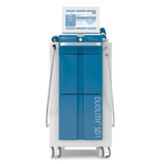
The first treatment was simple. The impacted tendon was treated with radial shockwave therapy (ESWT). The patient improved. Things seemed to be going well for about a month.
Then the pain came back.
When a patient has a recurrence, it is a setback for most patients. Clinically, it tells us something more valuable: where the original diagnosis was incomplete.
The findings extended well beyond the tibialis posterior tendon itself:
A successful initial response does not mean that the treatment is a failure if a recurrence occurs. It is a sign of diagnosis.
The question changed from “How do we treat this tendon?” to “Why does this tendon fail under load?”.
That’s the question that made all the difference.
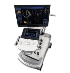
The clinical team went back to the medial hindfoot, but this time they used dynamic ultrasound evaluation instead of just static evaluations.
This is a very significant difference.
Static ultrasound reveals the structure of the tissue. Dynamic ultrasound reveals the behaviour of tissues when they are loaded and moved as opposed to reading a weather forecast and looking out the window.
The findings extended well beyond the tibialis posterior tendon itself:
| Structure Assessed | Finding |
|---|---|
| Tibialis posterior tendon | Tendinopathy confirmed |
| Talonavicular / spring ligament region | Abnormal opening detected |
| Tibio-spring portion of the deep deltoid–spring ligament complex | Abnormal appearance |
| Subtalar joint | Degenerative changes present |
It was no longer just a tendon issue.
The whole scenario was suggestive of overload of the tibialis posterior and a pattern of instability of the medial column, a very different clinical presentation and a very different treatment strategy.
To understand the diagnosis and why this distinction is important.
The main dynamic stabiliser of the medial arch is the tibialis posterior tendon. It is in constant use while walking, running and standing, it controls pronation and supports the arch with every step.
If the static stabilizers around it are lost (spring ligament, deep deltoid-spring ligament complex and talonavicular joint), the tibialis posterior tendon must take over the function of these stabilizers. It takes on loads for which it was not meant to be alone.
In this case, treating just the tendon is like changing a worn tire every time, but not fixing the wheel alignment that’s causing the wear.
The tendon might feel better for a short period of time. Though the mechanical environment that is causing it to be destroyed is unchanged.
That’s what makes the pain come back.
The treatment approach was restructured with a full picture now determined through dynamic ultrasound. Each of the elements addressed a specific part of the problem.
Prolotherapy was used for the spring ligament and deep deltoid-spring complex, which was found to have abnormal behavior during dynamic assessment.
In Prolotherapy, a specific irritating solution is injected into the body to trigger the body’s healing mechanisms in hypermobile or lax connective tissue. This was not a pain-killing exercise, but an exercise to restore integrity and mechanical stability of the ligaments.
The affected region of the joint was injected with Platelet-Rich Plasma (PRP).
PRP focuses the body’s own growth factors and targets them to degenerative or damaged tissue. In this case, it was the degenerative changes in the subtalar joint that were observed during dynamic assessment and would otherwise continue to aggravate the abnormal load distribution through the medial column.
Excessive medial column strain during the recovery period was reduced by using custom made temporary orthotics.
This is not a permanent solution. The purpose of orthotics here is to provide a specific functional role to protect the environment in which the prolotherapy and PRP can function without being immediately disturbed by the same forces that caused the injury.
It is as important to protect healing tissue as it is to treat it.
A focused extracorporeal shock wave therapy (ESWT) was repeated; yet there were significant differences in the targeting and intent.
Focused ESWT delivers energy to a particular depth and location, while radial shockwave spreads energy widely. This second application of shockwave therapy stimulated the deeper symptomatic structures which the first treatment had not stimulated enough.
This is an illustration of how the same treatment modality, with more comprehensive diagnostic information, yields a clinically different result.
After stabilization of the structural environment and management of the acute tissue response, the patient progressed to foot and ankle strengthening and progressive loading of the tibialis posterior.
Tendons need to be remodeled, strengthened and adapted through progressive mechanical load. By not loading the tendon at all during recovery, the tendon cannot regain its capacity. Though if the loading is too early, or too much in an unstable mechanical environment, the original injury pattern is simply reproduced.
The sequence stabilizes first then load is not a coincidence. It is the key to a successful result.
| Step | Intervention | Purpose |
|---|---|---|
| 1 | Dynamic ultrasound | Complete structural and mechanical diagnosis |
| 2 | Prolotherapy | Ligamentous stabilization |
| 3 | PRP | Joint and degenerative tissue repair |
| 4 | Temporary orthotics | Load protection during healing |
| 5 | Focused ESWT | Targeted stimulation of deeper structures |
| 6 | Progressive rehabilitation | Tendon reloading and functional restoration |
Each step creates the conditions that allow the next step to succeed.
This case is a good example of a principle that is applicable to many other tendon injuries.
Often pain is felt in one structure yet is the result of another.
The tissue that was injured was the tibialis posterior tendon. Yet the spring ligament, deep deltoid complex, talonavicular mechanics and subtalar joint were the culprits for the persistent pain.
The most common reason tendon treatments do not last is that only the painful structure is treated, but no attempt is made to understand why it is under excessive load.
Dynamic ultrasound enables clinicians to progress from a tissue-level diagnosis to a functional, mechanical diagnosis:
Not only “what is broken” but “why is it failing when it is put to the test?”.
This is the diagnostic standard for complex and recurrent musculoskeletal presentations.
We at NYDNRehab feel that it is important to report things truthfully as part of good clinical practice. The next section directly addresses the limitations inherent in this case study.
This is a single case report.
It’s about one patient, one clinical presentation and one treatment sequence. It is not a clinical trial and there was no control group or comparison treatment. The results should be viewed as an example of a clinical reasoning process rather than that this protocol leads to consistent results in all similar cases.
Several treatments were done at the same time.
The new treatment plan involved progressive rehabilitation, focused ESWT, orthotics, prolotherapy and PRP (all of which were added to the plan, but not all at once). This is often the case in complex clinical practice, where it may not be appropriate or ethical to wait to isolate variables.
But it does not mean that it is possible to know for sure which intervention had the greatest positive effect. It may be that orthotics alone were enough to get the load down to allow natural recovery. Improvement may have been due to rehabilitation alone. The truth is, we don’t know which part did the most, we know that it was when used in a logical sequence that it was able to solve dimensions of the problem that were not solved by the original treatment.
This report contains limited outcome data.
No formal pain scores, functional outcome measures, imaging follow-up or long term follow-up data are reported here. This is a real restriction. The next time such cases are reported at NYDNRehab, standardized outcome measures, including the Victorian Institute of Sport Assessment (VISA) score, visual analog pain scales, and follow-up ultrasound findings, will be included to better document treatment response.
There is varying evidence of individual treatments.
| Treatment | Current Evidence Level |
|---|---|
| Focused ESWT for tendinopathy | Moderate, supported by multiple RCTs |
| PRP for tendinopathy | Mixed, some positive RCTs, results vary by preparation and indication |
| Prolotherapy for ligamentous laxity | Emerging, promising but limited high-quality RCT data for foot ligaments specifically |
| Progressive tendon loading | Strong, well-established across tendinopathy research |
| Orthotics for tibialis posterior dysfunction | Moderate, supported for load management, less clear for long-term structural change |
Prolotherapy and PRP are both clinically promising and used in regenerative medicine, but have not been backed by as many controlled trial studies as shockwave therapy or exercise-based rehabilitation. Patients who are contemplating these procedures should have an informed discussion with their clinician about the current evidence base, realistic expectations and alternatives to these procedures.
Alternative explanations exist.
Initial shockwave therapy may have led to the return of pain for a variety of reasons other than medial column instability:
While the dynamic ultrasound findings gave a plausible structural explanation for the recurrence, there was not always a clear correlation between the imaging findings and the symptoms and the relationship between the imaging findings and the patient’s pain could not be said with absolute certainty.
The rationale behind the inclusion of this section.
This case isn’t so much about the proof of a protocol working, as it is about the value of the case itself. It’s because it shows a clinical reasoning process: how a recurrence can change the diagnostic question, how dynamic imaging can show mechanical contributors that static imaging does not, and how treatment strategy should be based on diagnosis, not treatment.
The following case is a good illustration of our thinking, but not a promise of results.
The shockwave therapy stimulates the healing process in the area, and it also helps to diminish the pain signals. If it provides temporary relief, but the symptoms come back, it usually indicates that the tendon has responded to treatment, but the mechanical factor that is causing it to be overloaded has not been corrected. The tendon heals, goes back to load and then fails again for the same reason. This reason was medial column instability.
The medial column is the inside of the foot, the arch, the talonavicular joint, the spring ligament and the ligament complexes around them. If these structures are weak or loose, the tibialis posterior tendon is called into action. That excess demand causes the tendon to wear over time. One of the most overlooked factors in tibialis posterior tendinopathy is medial column instability and one of the most important to diagnose and treat.
Radial shockwave therapy spreads energy out over a wide area, useful for diffuse or superficial soft tissue problems. Focused shockwave therapy applies energy to a specific area and depth, which is more suitable if the target structure is deeper or if imaging has pinpointed a location of pathology. Dynamic ultrasound in this instance provided clarity for which deeper structures required treatment, and thus focused ESWT was the more suitable modality for the second intervention.
Prolotherapy is a technique in which a concentrated solution (usually dextrose) is injected directly into the damaged or lax ligamentous tissue. The mild inflammatory response it triggers stimulates collagen production and gradual tightening of the ligament. It is especially beneficial for connective tissue that has become mechanically incompetent, but not fully torn, such as the spring ligament and deep deltoid-spring complex in this instance.
The tissue type and mechanism addressed by prolotherapy and PRP are different. Ligamentous laxity of support ligaments was treated with prolotherapy. PRP, a concentrated dose of the patient’s own growth factors, was targeted at the degenerative changes in the subtalar joint, which was not a ligament, but rather a joint. Both enabled the treatment to tackle a number of aspects of the same issue at once.
Not in this case. These orthotics were worn to minimize the strain on the medial column during the healing period, to prevent the reloading of the ligaments and joint tissue. The mechanical function of the orthotics was slowly phased out as the structural stabilizers regained their integrity and rehabilitation took place.
Dynamic ultrasound is particularly valuable when:
In many cases, yes. If the instability is ligamentous and not structural (bones are lined up but the soft tissue is not) then progressive rehabilitation and regenerative injections can restore meaningful mechanical stability. Surgery is indicated when the deformity is fixed, if conservative treatment has failed, and if the amount of joint degeneration is significant. No surgery was needed for this case.
If you have pain in your medial hindfoot or in your arch that doesn’t go away or gets better for awhile and then comes back, here are some questions to ask:
NYDNRehab is a specialty rehabilitation and sports medicine clinic in New York City known for its use of real-time dynamic ultrasound for diagnosis and treatment. The clinic’s philosophy of tendon, ligament and joint pathology is to look at the entire mechanical picture, not just the symptomatic tissue.
In more complicated, recurrent or previously treated musculoskeletal conditions, a more thorough diagnostic process is often the initial and most critical step in achieving long-term recovery.
Dr. Lev Kalika is a world-recognized expert in musculoskeletal medicine. with 20+ years of clinical experience in diagnostic musculoskeletal ultrasonography, rehabilitative sports medicine and conservative orthopedics. In addition to operating his clinical practice in Manhattan, he regularly publishes peer-reviewed research on ultrasound-guided therapies and procedures. He serves as a peer reviewer for Springer Nature.
Dr. Kalika is an esteemed member of multiple professional organizations, including:

Below is a prime example of how ultrasound can take the guesswork out of diagnosis.

A bad physical therapy experience is one of the primary causes of unnecessary surgery

In this instance, an athlete was originally diagnosed with minor quadriceps muscle strain and was treated for four weeks, with unsatisfactory results. When he came to our clinic, the muscle was not healing, and the patients’ muscle tissue had already begun to atrophy.
Upon examination using MSUS, we discovered that he had a full muscle thickness tear that had been overlooked by his previous provider. To mitigate damage and promote healing, surgery should have been performed immediately after the injury occurred. Because of misdiagnosis and inappropriate treatment, the patient now has permanent damage that cannot be corrected.
The most important advantage of Ultrasound over MRI imaging is its ability to zero in on the symptomatic region and obtain imaging, with active participation and feedback from the patient. Using dynamic MSUS, we can see what happens when patients contract their muscles, something that cannot be done with MRI. From a diagnostic perspective, this interaction is invaluable.
Dynamic ultrasonography examination demonstrating
the full thickness tear and already occurring muscle atrophy
due to misdiagnosis and not referring the patient
to proper diagnostic workup
Demonstration of how very small muscle defect is made and revealed
to be a complete tear with muscle contraction
under diagnostic sonography (not possible with MRI)
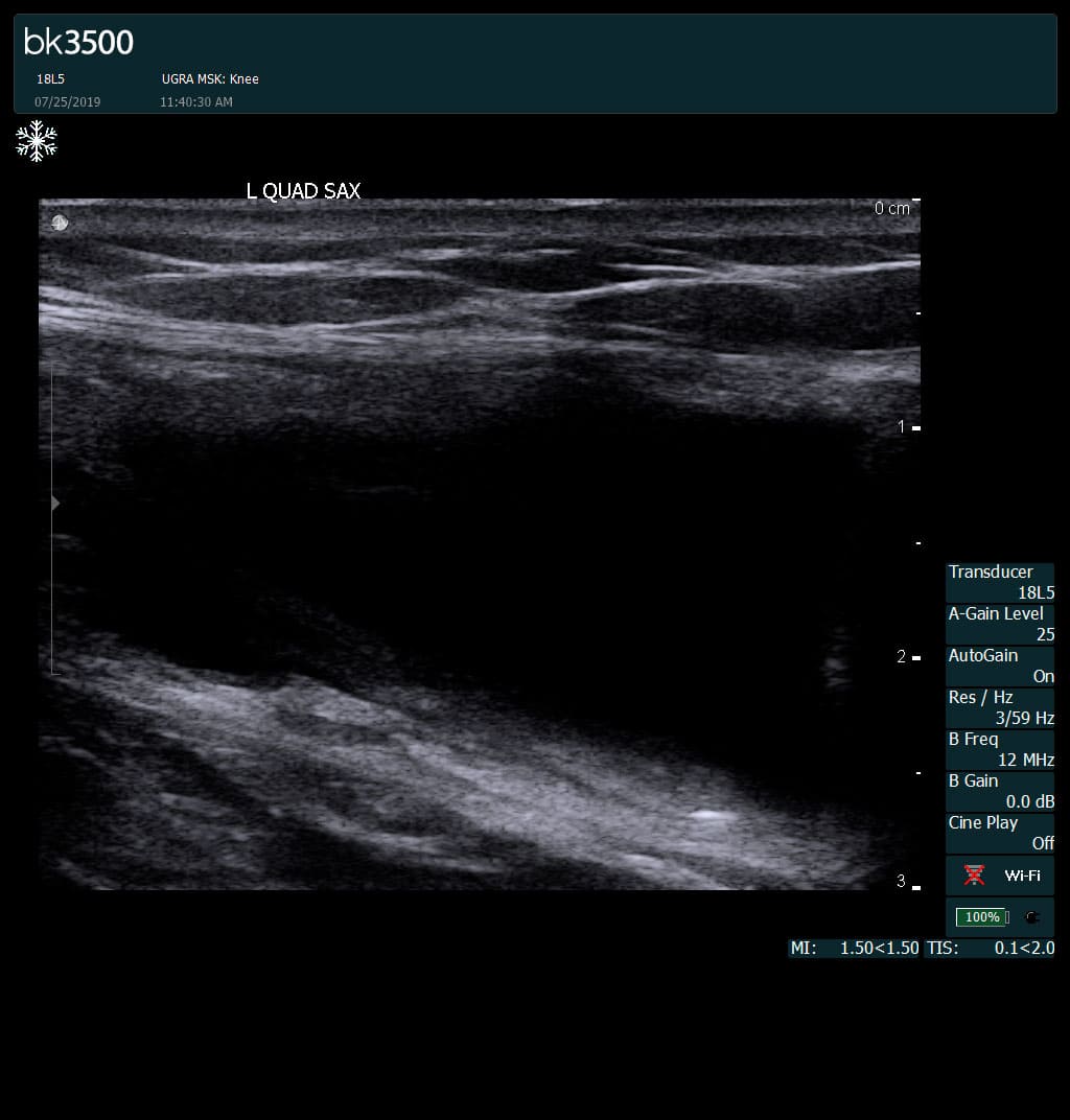
Complete tear of rectus femoris
with large hematoma (blood)
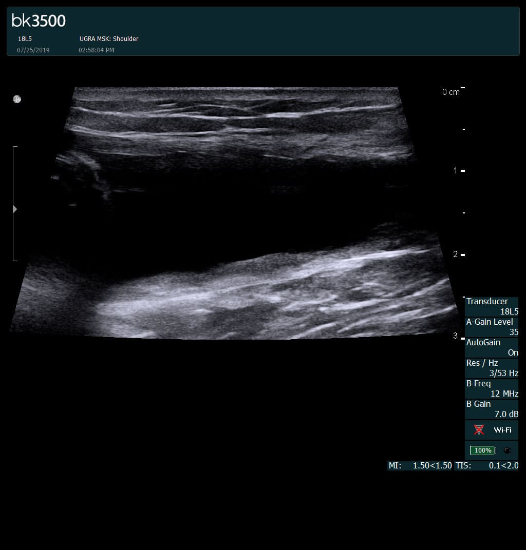
Separation of muscle ends due to tear elicited
on dynamic sonography examination


 Request Telehealth