Diagnostic examination and treatment performed by Dr. Lev Kalika, DC, RMSK, clinical director of NYDNRehab, registered in musculoskeletal sonography (RMSK, ARDMS/Inteleos) since 2012.

A patient who had undergone surgery for a dorsal wrist ganglion cyst had been complaining of persistent dorsal wrist pain for two years.
During that time she saw four hand surgeons. Repeatedly reassured that the wrist looked fine on routine MRI and no significant abnormality was seen. The pain did not change, and it continued to limit her daily activities.
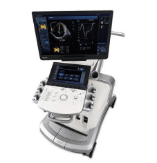
High-resolution musculoskeletal ultrasound of the wrist was repeated, with the contralateral side being compared to the asymptomatic side in the same examination.
The symptomatic wrist had:
The sonographic findings were located in the same place and behaved in the same way as her symptoms.

Treatment combined two ultrasound-guided approaches. The first was hydrodissection of the scar from the surgery. The injectate was placed in the interfascial plane between the extensor tendons and the dorsal capsule, separating the adherent layers to restore glide. The term is used for the procedure that is performed around the median nerve in carpal tunnel syndrome, but the target is different.
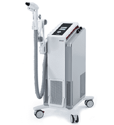
The second was a course of directed focused extracorporeal shockwave therapy (fESWT), which focused on improving the mobility of the tissue plane opened by the hydrodissection and remodeling the scar.
This patient felt that symptoms resolved completely and functioned normally without pain.
For most wrist disorders, routine MRI is a good modality. Resolution wasn’t the limiting factor in this case. It was the absence of motion.
A small superficial scar may be inconspicuous on the still frame but may bind the tendons that cross it. That binding only appears when the tendons are called upon to move. The published literature on postoperative imaging of the hand and wrist makes the same distinction; static imaging will demonstrate the presence of scar tissue, but without dynamic assessment it cannot establish how much that scar restricts tendon excursion. Ultrasound has an established role in detecting tendon adhesions and other local complications after hand and wrist surgery.
High-resolution musculoskeletal ultrasound adds the following:
This is the same series of dynamic diagnosis and ultrasound guided scar release that has been reported for tendon tethering at other sites. The effects of shockwave therapy on extracellular matrix remodeling in dermal and subdermal fibrosis have been documented, and the signaling pathways involved have been reviewed systematically.
The finding was never hidden. It needed a modality that would be able to follow the movement of the tissue.
One of the most frequent complaints of patients who visit NYDNRehab is persistent pain following wrist surgery where there is no remarkable imaging of the wrist. The difference between the images and the symptoms is often explained by a dynamic ultrasound examination with side-to-side comparison. Please contact the clinic to make an appointment.
All cases described on this site are evaluated and managed by Dr. Lev Kalika and the NYDNRehab clinical team.
Dr. Lev Kalika is a world-recognized expert in musculoskeletal medicine. with 20+ years of clinical experience in diagnostic musculoskeletal ultrasonography, rehabilitative sports medicine and conservative orthopedics. In addition to operating his clinical practice in Manhattan, he regularly publishes peer-reviewed research on ultrasound-guided therapies and procedures. He serves as a peer reviewer for Springer Nature.
Dr. Kalika is an esteemed member of multiple professional organizations, including:

Below is a prime example of how ultrasound can take the guesswork out of diagnosis.

A bad physical therapy experience is one of the primary causes of unnecessary surgery

In this instance, an athlete was originally diagnosed with minor quadriceps muscle strain and was treated for four weeks, with unsatisfactory results. When he came to our clinic, the muscle was not healing, and the patients’ muscle tissue had already begun to atrophy.
Upon examination using MSUS, we discovered that he had a full muscle thickness tear that had been overlooked by his previous provider. To mitigate damage and promote healing, surgery should have been performed immediately after the injury occurred. Because of misdiagnosis and inappropriate treatment, the patient now has permanent damage that cannot be corrected.
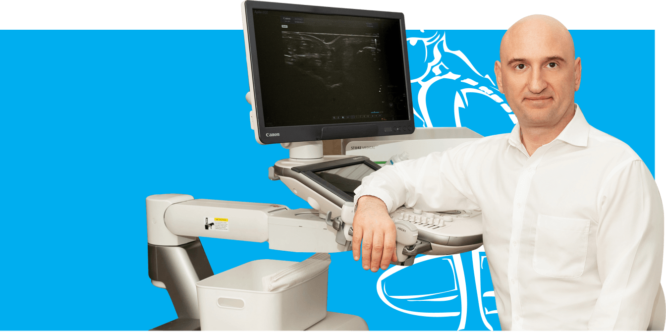
The most important advantage of Ultrasound over MRI imaging is its ability to zero in on the symptomatic region and obtain imaging, with active participation and feedback from the patient. Using dynamic MSUS, we can see what happens when patients contract their muscles, something that cannot be done with MRI. From a diagnostic perspective, this interaction is invaluable.
Dynamic ultrasonography examination demonstrating
the full thickness tear and already occurring muscle atrophy
due to misdiagnosis and not referring the patient
to proper diagnostic workup
Demonstration of how very small muscle defect is made and revealed
to be a complete tear with muscle contraction
under diagnostic sonography (not possible with MRI)
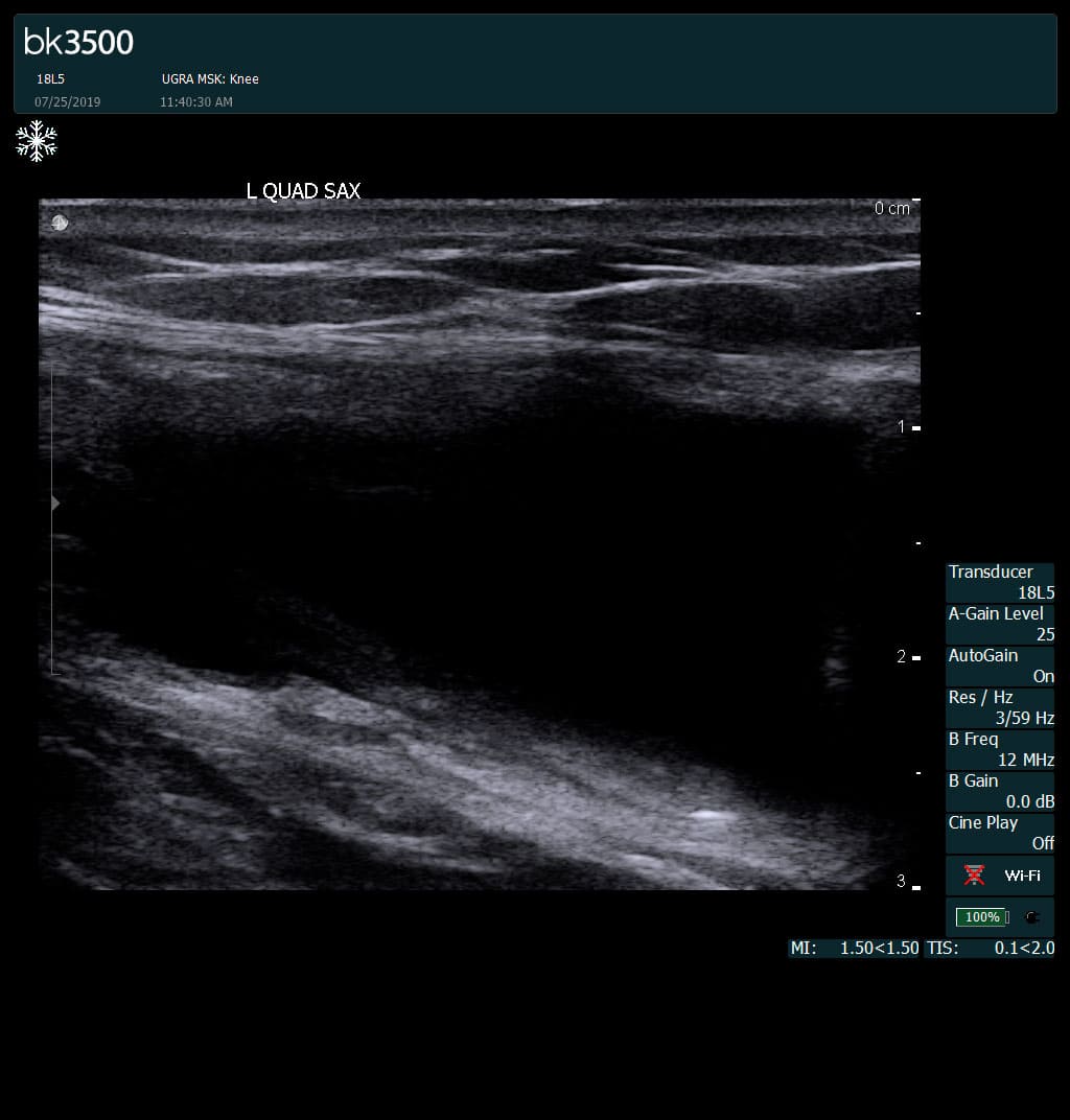
Complete tear of rectus femoris
with large hematoma (blood)
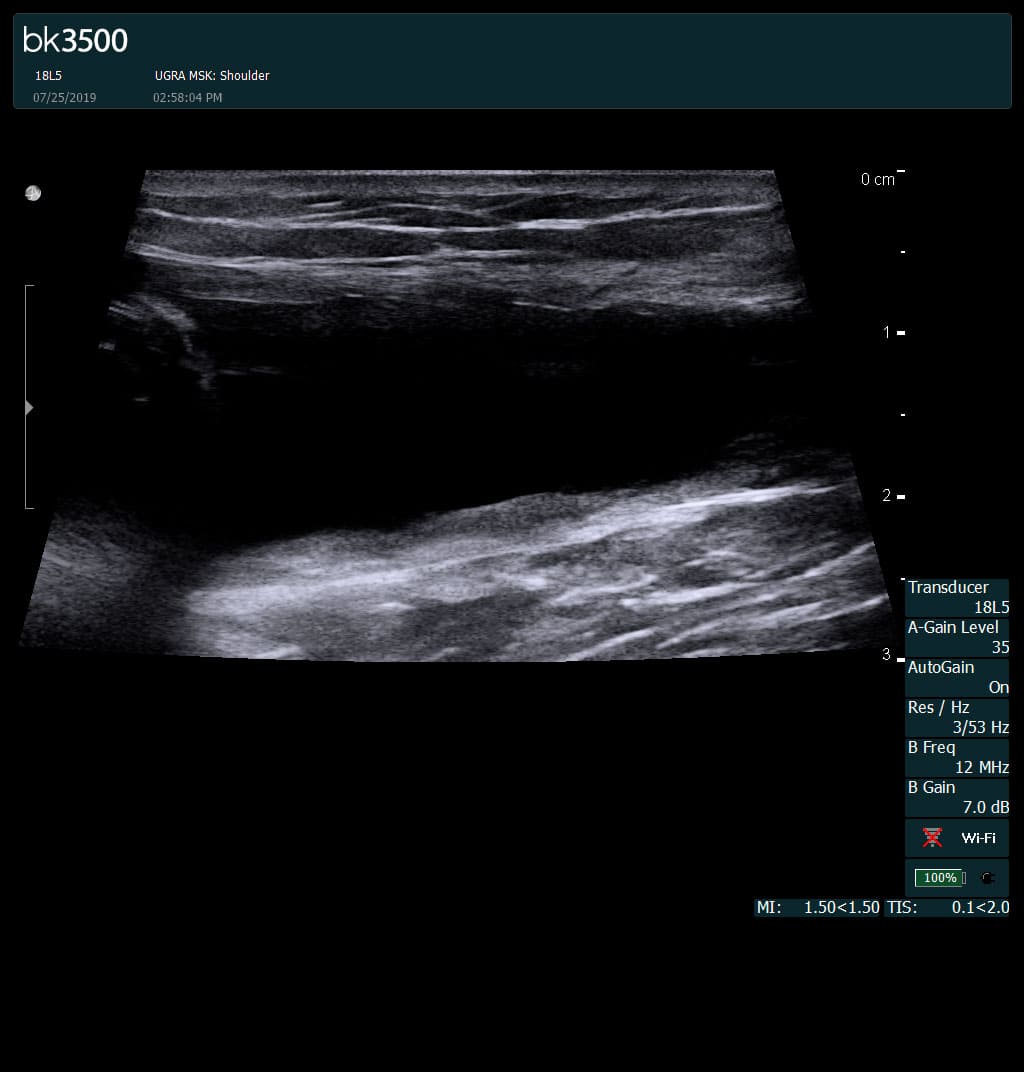
Separation of muscle ends due to tear elicited
on dynamic sonography examination


 Request Telehealth