A 51-year-old woman came to us after being treated with physical therapy for “shoulder impingement.” She was not improving. If anything, she was getting worse.
When I examined her shoulder with high-resolution musculoskeletal ultrasound, the first abnormality was obvious: a relatively soft calcific deposit within the supraspinatus tendon. That could easily have been the end of the story.
But when I looked carefully at the tendon insertion, something else caught my attention. The deep fibers near the footprint almost looked like a partial articular-sided tear. Almost. Something did not fit. So instead of labeling it, I traced the supraspinatus fibers in real time, from tendon to footprint and back again.
That changed the diagnosis. What initially resembled a partial-thickness tear was actually cortical erosion of the greater tuberosity with intraosseous extension of the calcific deposit. This is an uncommon but important manifestation of calcific tendinopathy: during the resorptive phase, calcium can become less dense and migrate out of its original location, most often toward the bursa, but occasionally into bone or even muscle.
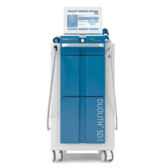
This is where diagnosis becomes treatment. There were two reasonable minimally invasive options to consider. Ultrasound-guided barbotage mechanically needles, irrigates and aspirates the calcific material. The alternative is focused extracorporeal shockwave therapy (fESWT).
In this particular case, I strongly preferred fESWT. The reason is that I was no longer treating only a calcification inside a tendon. There were two involved tissues: the supraspinatus tendon and the underlying bone. Focused shockwave has a well-established role in calcific rotator-cuff tendinopathy and can promote resorption of calcific deposits, and focused shockwaves are also used in bone pathology, where mechanotransduction can stimulate vascular and bone-remodeling responses.
So rather than passing a needle through an already disrupted tendon-bone interface to remove a soft deposit, fESWT gave us the opportunity to address both sides of the lesion, the calcification and the bone response. A potential double win. This also fits what the comparative literature suggests: once a deposit has eroded into bone, it is often no longer a straightforward target for percutaneous irrigation.
This case illustrates something more important than a naming error. Every diagnosis creates a treatment strategy. If we diagnose “impingement,” we may repeatedly strengthen and load the shoulder as an impingement problem. But if the real problem is an active resorptive calcific tendinopathy with cortical erosion and intraosseous involvement, repeatedly loading a highly reactive tendon-bone interface may be exactly what the patient does not need at that stage.
The physical therapy itself was not necessarily wrong. It was being applied to the wrong diagnosis. That distinction matters.
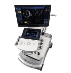
At NYDNRehab, this is why we place so much emphasis on precision diagnosis before treatment. Musculoskeletal ultrasound is not simply about finding something abnormal. It is about understanding what tissue is involved, what biological process is occurring, and therefore what treatment actually makes sense. Sometimes the most important part of an ultrasound examination is knowing when not to stop looking.
If you have shoulder pain that is not responding to treatment, a precise diagnosis is the first step. You can contact our clinic to arrange an assessment.

Dr. Lev Kalika is a world-recognized expert in musculoskeletal medicine. with 20+ years of clinical experience in diagnostic musculoskeletal ultrasonography, rehabilitative sports medicine and conservative orthopedics. In addition to operating his clinical practice in Manhattan, he regularly publishes peer-reviewed research on ultrasound-guided therapies and procedures. He serves as a peer reviewer for Springer Nature.
Dr. Kalika is an esteemed member of multiple professional organizations, including:

Below is a prime example of how ultrasound can take the guesswork out of diagnosis.

A bad physical therapy experience is one of the primary causes of unnecessary surgery

In this instance, an athlete was originally diagnosed with minor quadriceps muscle strain and was treated for four weeks, with unsatisfactory results. When he came to our clinic, the muscle was not healing, and the patients’ muscle tissue had already begun to atrophy.
Upon examination using MSUS, we discovered that he had a full muscle thickness tear that had been overlooked by his previous provider. To mitigate damage and promote healing, surgery should have been performed immediately after the injury occurred. Because of misdiagnosis and inappropriate treatment, the patient now has permanent damage that cannot be corrected.
The most important advantage of Ultrasound over MRI imaging is its ability to zero in on the symptomatic region and obtain imaging, with active participation and feedback from the patient. Using dynamic MSUS, we can see what happens when patients contract their muscles, something that cannot be done with MRI. From a diagnostic perspective, this interaction is invaluable.
Dynamic ultrasonography examination demonstrating
the full thickness tear and already occurring muscle atrophy
due to misdiagnosis and not referring the patient
to proper diagnostic workup
Demonstration of how very small muscle defect is made and revealed
to be a complete tear with muscle contraction
under diagnostic sonography (not possible with MRI)
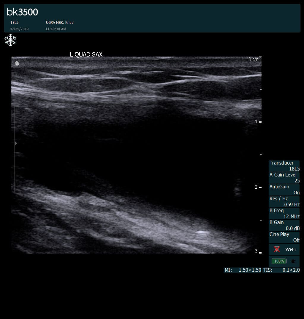
Complete tear of rectus femoris
with large hematoma (blood)
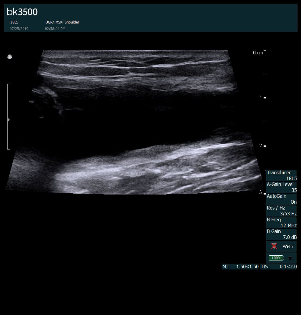
Separation of muscle ends due to tear elicited
on dynamic sonography examination


 Request Telehealth