Chronic pain is a problem that plagues millions of people and for many, surgery is finally considered as a solution. That conversation is sometimes months after a failed treatment. It can occur quite early on. In either case, when most patients come to this point, they don’t have a good idea of what surgery can actually achieve or what it can’t.
This article discusses 10 common misunderstandings about surgery for chronic musculoskeletal pain. It’s not an argument against surgery. There are some operations that are indeed needed, evidence-based and life-changing for the appropriate patient. It really does matter if you’re the “right patient” or not, and there’s not enough time in most pre-surgical consultations for it to be a big concern.
The truth is, it depends on the nature of the “conservative treatment”.
According to a 2024 global comparison of clinical practice guidelines published in BMC Musculoskeletal Disorders, the majority of musculoskeletal conditions including chronic back pain, knee osteoarthritis, rotator cuff problems and tendinopathy call for a true trial of non-surgical treatment before surgery is considered. This includes structured physiotherapy, progressive exercise, load management and sometimes injection therapy and pain education. Here, the term structured is important. A few exercises not tailored to your issue are not the same as a well-designed rehab program.
Patients often come to a surgical consultation with a treatment they’ve been on that was too short, too vague or, simply, the wrong treatment. It’s not a sign of failure of conservative care, it’s a sign that a specific strategy was not a fit for a given problem. Before deciding that surgery is the next step, it’s important to know if the causes of your pain have been determined and treated.
Questions to ask: Why am I feeling pain? (Not where am I feeling pain!) Was your rehabilitation program progressed and modified over time or was it the same program of exercises throughout?


The truth: Surgery corrects structure. It doesn’t always cure pain, and the two are not synonymous.
There are more cases of chronic pain following a technically successful procedure than is commonly discussed prior to surgery. One of the primary reasons is a phenomenon known as “central sensitization”, which occurs when the nervous system becomes overactive as a result of prolonged exposure to pain.
So, if pain has been around for months or years, the brain and spinal cord can become stuck in an “on” state. The nervous system has re-calibrated around the pain and pain signals continue to fire after the initial tissue problem is solved. The structure is repaired with surgery. It can’t recalibrate the nervous system.
When central sensitization is a major contributor to the problem, then results from surgery are often disappointing, not because the surgery was poorly conducted, but because the surgery was never going to be able to solve the real problem.
This is one of the reasons psychological assessment before surgery and pain neuroscience education are gaining traction as clinically relevant interventions, rather than a formality.
Takeaway: If you have been suffering from a great deal of pain for a long period of time, ask your surgeon or pain specialist if they have considered central sensitization and if it would alter your treatment options.


The truth: The connection between structural changes and pain is far less than what most think.
Studies have repeatedly demonstrated that the same types of findings (such as thinning cartilage, meniscal changes, and disc degeneration) are seen in people with and without symptoms. A popular imaging study of healthy adults revealed that most people had some level of disc deterioration on MRI, but not a single one was experiencing symptoms. This is also seen in studies of the shoulder, knee and hip.
This is not to say that structural degeneration is unimportant. It does imply, however, that it is a big mistake to think that the pain is caused by something that is visible, and that getting rid of or replacing that something will cure the pain.
The experience of pain is modified by factors such as body weight, sleep quality, physical conditioning, systemic inflammation and psychological stress. Near identical imaging can result in vastly different pain levels, and that’s where many failed surgeries reside.
The key takeaway: If there is an abnormality on the scan, this is what your joint looks like. Does not always describe the cause of pain.


The truth: For many patients, osteoarthritis progression is more controllable than they’ve been led to believe.
Body weight, metabolic health and physical activity are strongly associated with OA, and these are factors that are potentially modifiable. A 2024 systematic review and meta-analysis in Arthritis Care & Research confirmed consistently strong effects of exercise specifically appropriately loaded resistance training) on pain and physical function in knee and hip OA. The less weight that goes through a joint, the less mechanical stress it receives with each step, and even small decreases can make a significant difference in symptom improvement.
Other treatments known as orthobiologics, which involve the use of components found in the patient’s own blood to stimulate tissue repair, are also being studied and applied in the clinic. The evidence base is emerging and is different for each treatment and condition, but for some patients with OA in the early stages, these methods could help to delay or minimise the need for replacement surgery. https://www.aaos.org/globalassets/quality-and-practice-resources/biologics/technology-overview_prp-for-knee-oa.pdf
For appropriate candidates, joint replacement is a well established procedure and has a good success rate. The problem is not that it doesn’t work, it’s that sometimes it is made to look like it is inevitable when it’s not.
Questions to ask: Have you participated in an exercise program under supervision for OA? Have there been any talks about weight management? Are there alternatives to surgery that can be tried first?


The truth: Spinal fusion is a spine procedure that has a definite place in spine care, but most cases of chronic low back pain do not fit that mold.
Spinal fusion is a surgery that permanently fuses two or more vertebrae together, so that they are no longer able to move. It is suitable for situations where there is a true structural instability (spondylolisthesis, some fractures, some cases of severe nerve compression). In the case of non-specific chronic low back pain (pain without a specific structural cause), the evidence is less supportive.
There are several well-designed trials that have compared surgical fusion with structured rehabilitation for non-specific chronic back pain, with no long-term benefit of surgery. Fusion, on the other hand, has real risks: infection, hardware failure and adjacent segment disease, which is the progressive deterioration of the spinal segments above or below the fused segment, with increased mechanical stress once the fused segment has been locked.
There are also many factors that surgery can’t fix that contribute to back pain like movement patterns, muscle imbalances, load habits, psychological stress. They are not visible on imaging and can’t be fixed in theatre.
Questions to ask: What is the particular structural defect that makes me a surgical candidate? What are the facts regarding fusion in people with my diagnosis? What will be the fate of the segments over and under the fusion with the passage of time?


The reality: ACL reconstruction is a good procedure for some patients but not for all.
In fact, according to Jia et al. in their comprehensive analysis published in Journal of Orthopaedics (2024), the functional recovery of a patient is pretty much the same after undergoing surgery or a strict, non-operative rehab treatment plan. The outcomes of surgery seemed to be moderately good for short-term knee stability, but there were fewer significant improvements in knee function over long-term follow up.
Surgical intervention remains most relevant for athletes aiming for a swift return to professional sports and for patients with persistent instability which has a major impact on their daily activities. A specialized non-surgical approach may have similar results in certain populations, such as older adults or people who do not use pivoting motion in their activities of daily living. Also, some studies have cast doubt on the long-term health of cartilage following reconstruction, with some studies reporting higher rates of knee osteoarthritis. This is still an ongoing research.
Important: The ACL tear is a significant injury but not one that has to be repaired. The right choice will vary according to the age, activity level, amount of instability and involvement of other structures.


The truth: This is perhaps the most clinically relevant myth on this list, patients often don’t realize that there’s a big gap between “there’s a tear on the scan” and “you need surgery.” What MRI shows isn’t always what it seems.
Tears of the meniscus, or knee cartilage, are frequent. So is the inability to find them in individuals that do not have any significant knee symptoms. Though there is another aspect to this: not all MRI findings of a tear are a true tear.
A meniscal flounce, which is a normal wavy or folded appearance of the inner free edge of the meniscus, can appear on imaging like a tear, especially if only a single MRI slice is analyzed. One of the classic imaging pitfalls in knee MRI and has absolutely no clinical significance.
Other normal anatomical structures commonly mistaken for tears are:
The real-world implication: A radiologist’s report that says you have a tear should always be read in conjunction with your symptoms and examination, and how the injury occurred, and not on its own.
When surgery is genuinely indicated:
There are certain tear patterns where surgery is clearly appropriate and sometimes urgent. The most obvious is locked knee due to a displaced bucket handle tear. In this case, a piece of meniscus has become dislodged and is blocking the knee from straightening. This can’t be fixed with rest and rehabilitation, and delay can cause cartilage damage. Usually surgery is advised early.
In other cases, surgery is the best option:
When conservative care comes first:
Degenerative horizontal cleavage tears, small stable tears, MRI findings without true mechanical symptoms, and tears in knees with advanced arthritis are generally managed conservatively first – rehabilitation, load modification, and sometimes injection therapy — with surgery considered only if meaningful mechanical symptoms persist. Several randomised trials, including some that employed sham surgery as a control group, have supported this approach for degenerative tears in middle-aged adults.
Questions to ask: What kind of tear is it, traumatic or degenerative? Is there a mechanical block or is it just pain? Does my tear pattern fit the profile where surgery has the strongest evidence of benefit?


The reality: Surgery is meaningfully safer than it was a generation ago. That’s not the same as low-risk.
Even technically well-executed surgery can produce irreversible changes that outlast the original problem. Arthrofibrosis, scar tissue formation, incidental nerve or vascular damage, and disruption to the body’s fascial system are all recognised, if underacknowledged, risks of surgical intervention. In all operations the fascia network of continuous connective tissue is inevitably divided and remodeled. Restrictions in fascial sliding ability can take on the quality of a spreading effect and affect pain and movement throughout the body through ‘fascial chains’, away from the site of surgery. This is less often discussed in the pre-operative consultation and it is a part of the reason for the lack of focus on fascial preservation in traditional orthopaedic training. The structure of the body that comes out of the operating theatre is reorganized and the downstream effects of that reorganization are not always predictable, visible on imaging, or reversible. https://pubmed.ncbi.nlm.nih.gov/18789982/
The outcomes can also be influenced by the type of procedure, the health of the patient, the skill of the surgeon, and rehabilitation following the surgery. An average performing procedure in a clinical trial will yield some very different outcomes in an individual scenario. None of this should be used as an excuse not to do the surgery when it is truly indicated; it should be an excuse to consider carefully and slowly any surgery recommendation.


The reality: Long periods of bed rest are ineffective, and can sometimes prove detrimental, for most orthopaedic surgeries.
Within days muscles begin to lose mass when not used. It also decreases blood flow and makes it more likely that the blood will clot. It makes it difficult to get better because it causes changes in the connective tissue and makes the joints stiff. Early mobilisation (within 24–48 hours after surgery, when clinically appropriate) is beneficial in terms of a number of evidence-based measures, including: reduced hospital stay, decreased complications, better pain management, and earlier return to function.
If that is what you believe is best for your area, then it is understandable. But your physical therapist will want you moving at a much earlier time than most people would think and for good reason.


The reality: The weeks before surgery are more important than what patients are told!
There is a good evidence base for prehabilitation, which is a structured programme of exercise, optimisation of nutrition and mental preparation before an operation. As outlined in a systematic review in the Journal of Surgical Research by Kann et al., 2025, patients undergoing prehabilitation had superior early functional outcomes and shorter hospital lengths of stay than those who did not receive prehabilitation.
Increasing muscle around the affected joint, exercising to increase cardiovascular fitness, correcting nutritional deficiencies, optimizing sleep patterns, and reducing pre-operative anxiety seems to be all aiding in the body’s response to surgical stress and efficient recovery. The idea is simple: the stronger and better prepared the body the better it will do in the operation.
Takeaway: If your surgery is in the future, inquire about prehabilitation. A little preparation can make a big difference in just a few weeks.


Yet, there are also a number of newer, more specialised, non-surgical treatments that are now used in the treatment of chronic musculoskeletal issues, particularly where other treatments have failed or where patients prefer not to have or delay surgery.
These include:
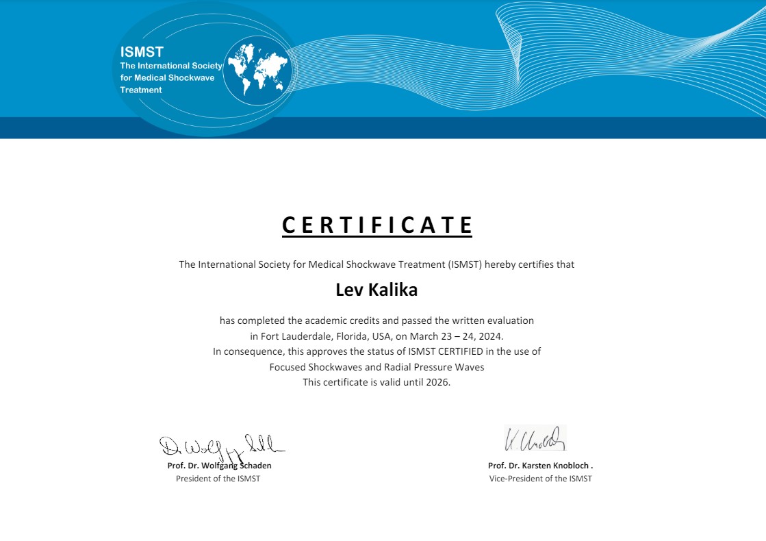
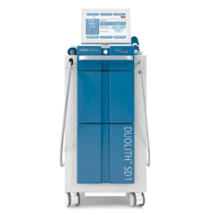
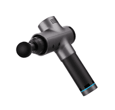
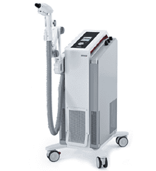
Extracorporeal Shockwave Therapy (ESWT)
Shoots focused sound waves into the tissue to encourage healing and stop pain. Evidence base for chronic tendinopathy, plantar fasciitis and shoulder calcific tendinitis is reasonable.
It’s also important to know how shockwave therapy fits into the process of post-surgical rehabilitation. When and how it’s used is a lot more important than many patients think. Shockwave therapy should be applied sparingly and sensibly in the early post-operative period to avoid employing it as a “violent” healing accelerator. The key to this stage is to minimize pain, swelling, maximize the microcirculation and prevent excessive stiffness or adhesion of the fascia while always respecting the surgeon’s healing phase.
Early application will often be directed around the surgical area instead of through the area. Not suitable targets include fresh repairs, incisions, hardware, graft fixation points, or newly sutured tendon or ligament tissue. A low energy radial or defocused shockwave can be applied to surrounding soft tissues to help reduce protective muscle spasm, promote lymphatic drainage and restore tissue glide, without over stimulating the vulnerable structures. Focused shockwave, which can go deeper and pack more energy, is typically used when the repair is not biologically stable (usually in the context of chronic enthesopathy, delayed tendon-bone healing or focal scar restriction that has not improved on its own).
The rule of thumb is simple – the shockwave should be therapeutic, not traumatic, during the early stages following surgery. All matters of dose, timing and precision. With proper use and careful loading, mobility activities and swelling control, it can contribute to improved motion and comfort for patients. It can cause more damage than good if not used properly, too soon, too heavily, or if it is not used for what was corrected during surgery.
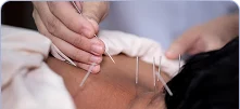
Percutaneous Electrical Nerve Stimulation (PENS)
Is the one method that is currently being used.
Fine needle electrodes are inserted into the body near the affected nerve and low frequency electrical stimulation is applied to decrease the hypersensitivity. Suitable if the nervous system is believed to be an important contributor to chronic pain.
Prolotherapy
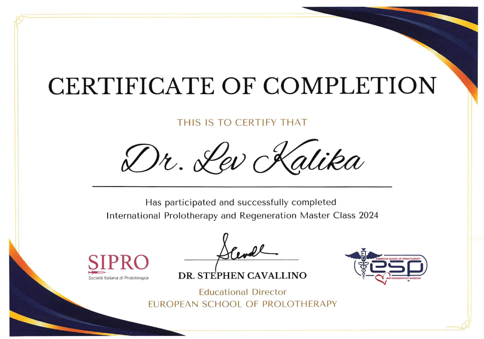
A technique that uses a mild irritant solution to stimulate the body’s natural healing process by injecting it into damaged areas of ligament or tendon tissue. It has been used for decades for a number of conditions including chronic low back pain, ligament laxity, and others, with evidence base varying by condition.


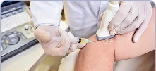
This involves the injection of saline solution into the fascial layers or around nerve entrapment under ultrasound guidance to help loosen adhesions and alleviate nerve entrapment. It’s a carefully controlled, minimally invasive procedure that can be performed in various situations, such as carpal tunnel syndrome or some instances of sciatic nerve irritation.
Fascial Manipulation (Stecco Method)
A manual therapy technique, which is hands-on, focuses on areas of fascia that are both swollen and tight (the connective tissue that runs throughout the body) and aims to free up normal range of freedom and relieve pain.
None of these are ‘one size fits all’ solutions and not all will be relevant to every patient and to every condition. But they’re not just a limited choice, as many patients are not aware of all the options at the time their surgeon mentions they may need surgery.

If surgery has been recommended and you are unsure, there are a couple of things that you should do. Discuss with your surgeon, in detail, why surgery is recommended for your condition, what you can reasonably expect from surgery in terms of pain relief (which may not be complete), and what options are available if you do not have surgery. If in doubt, get a second opinion. And ensure that conservative measures have been seriously considered, not superficially tried.
Chronic pain is complex, there are no universal solutions. Knowing what surgery can and cannot achieve, and what other choices are available, will greatly help you make a decision that’s right for your situation.
Request an appointment»

Our location: 11 West 25th Street 5th floor, New York, NY 10010
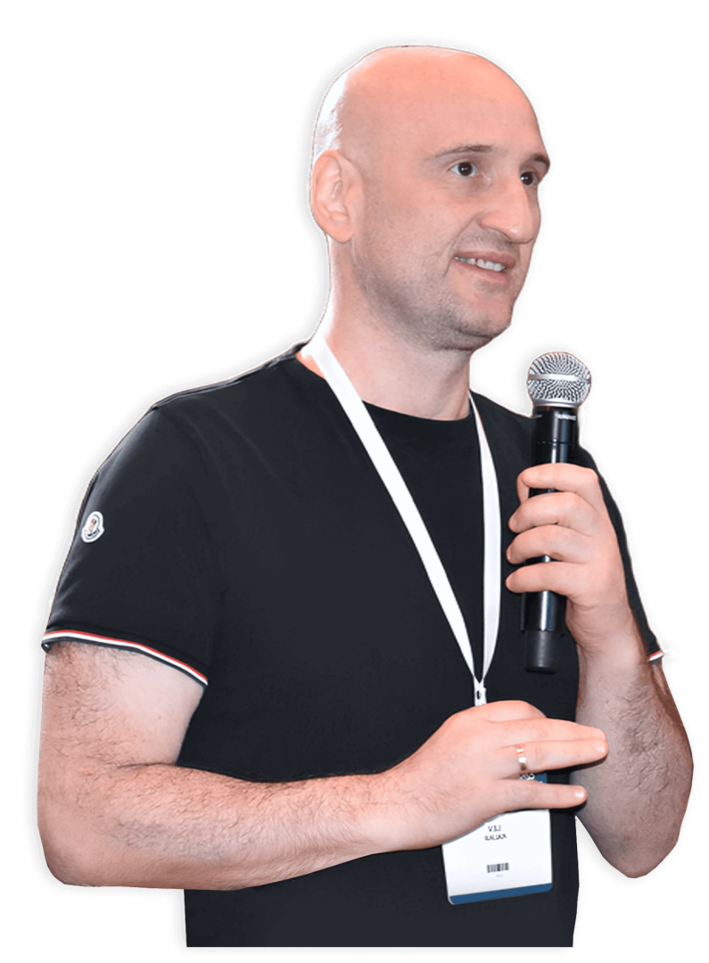
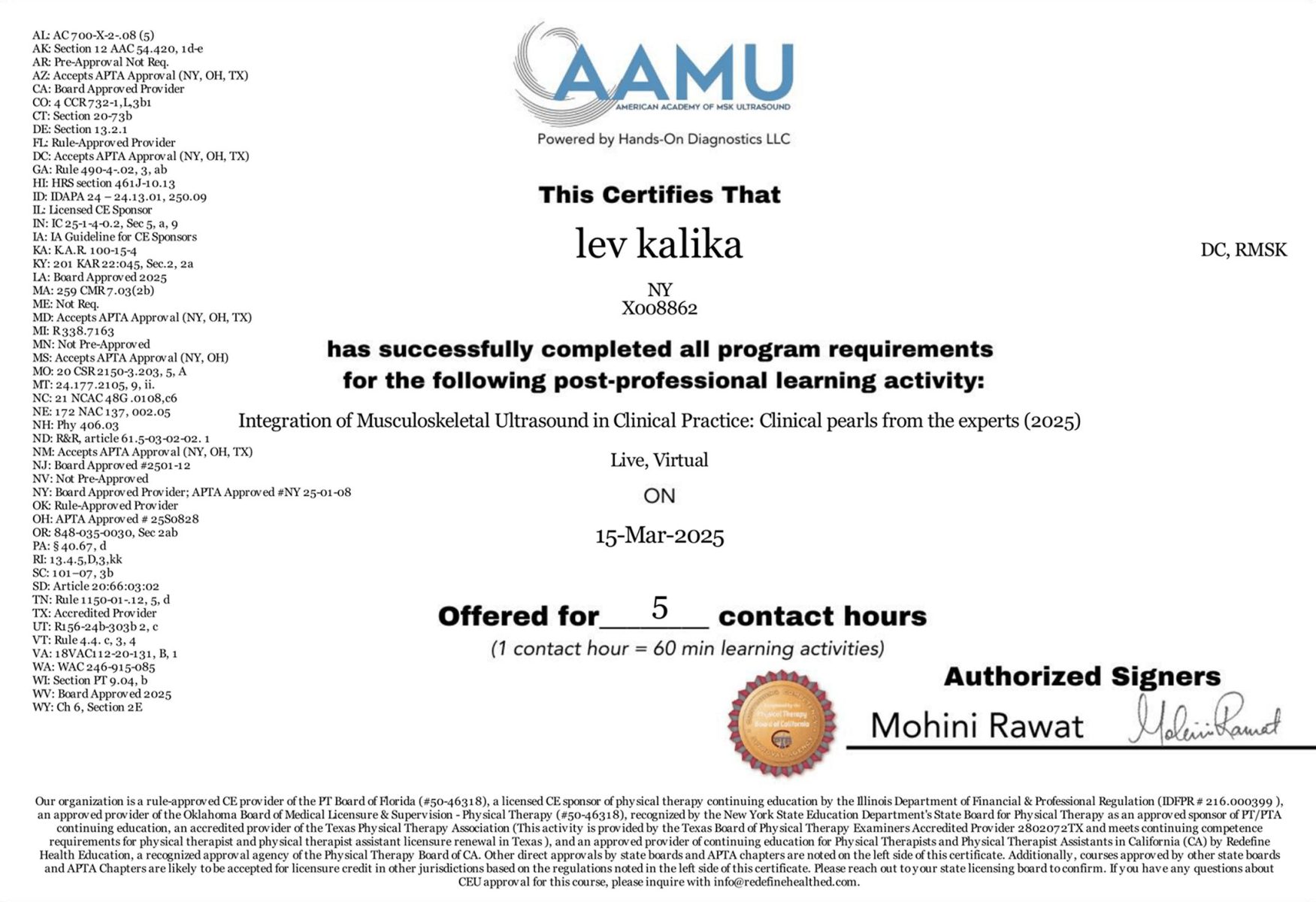
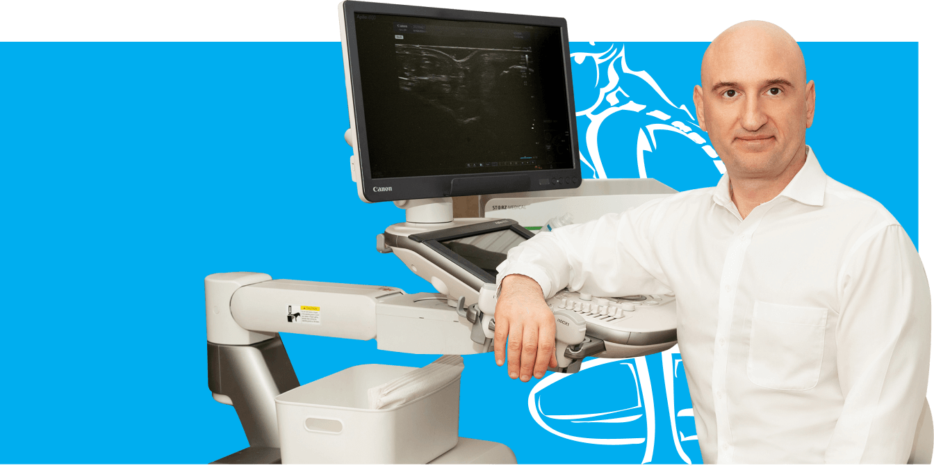
Dr. Lev Kalika is a world-recognized expert in musculoskeletal medicine. with 20+ years of clinical experience in diagnostic musculoskeletal ultrasonography, rehabilitative sports medicine and conservative orthopedics. In addition to operating his clinical practice in Manhattan, he regularly publishes peer-reviewed research on ultrasound-guided therapies and procedures. He serves as a peer reviewer for Springer Nature.
Dr. Kalika is an esteemed member of multiple professional organizations, including:

Below is a prime example of how ultrasound can take the guesswork out of diagnosis.

A bad physical therapy experience is one of the primary causes of unnecessary surgery

In this instance, an athlete was originally diagnosed with minor quadriceps muscle strain and was treated for four weeks, with unsatisfactory results. When he came to our clinic, the muscle was not healing, and the patients’ muscle tissue had already begun to atrophy.
Upon examination using MSUS, we discovered that he had a full muscle thickness tear that had been overlooked by his previous provider. To mitigate damage and promote healing, surgery should have been performed immediately after the injury occurred. Because of misdiagnosis and inappropriate treatment, the patient now has permanent damage that cannot be corrected.
The most important advantage of Ultrasound over MRI imaging is its ability to zero in on the symptomatic region and obtain imaging, with active participation and feedback from the patient. Using dynamic MSUS, we can see what happens when patients contract their muscles, something that cannot be done with MRI. From a diagnostic perspective, this interaction is invaluable.
Dynamic ultrasonography examination demonstrating
the full thickness tear and already occurring muscle atrophy
due to misdiagnosis and not referring the patient
to proper diagnostic workup
Demonstration of how very small muscle defect is made and revealed
to be a complete tear with muscle contraction
under diagnostic sonography (not possible with MRI)
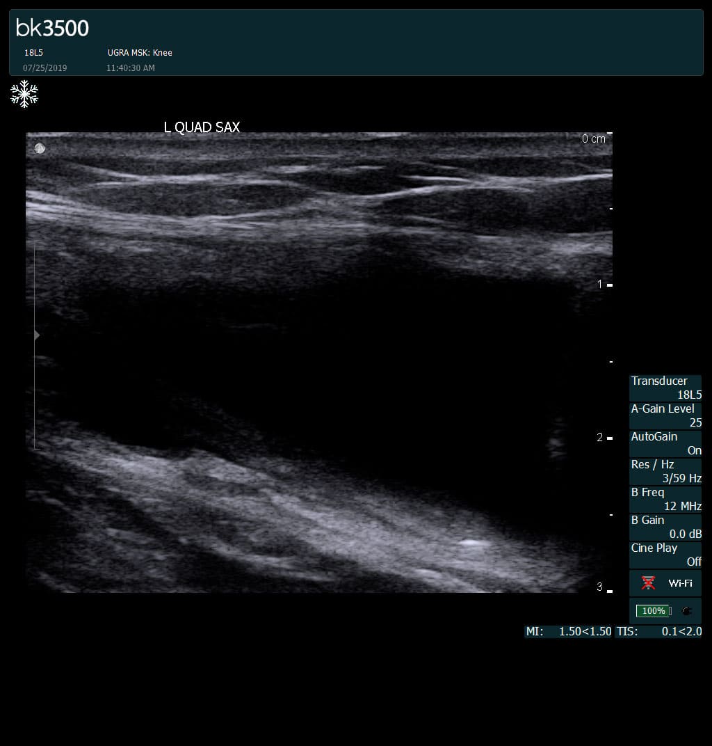
Complete tear of rectus femoris
with large hematoma (blood)
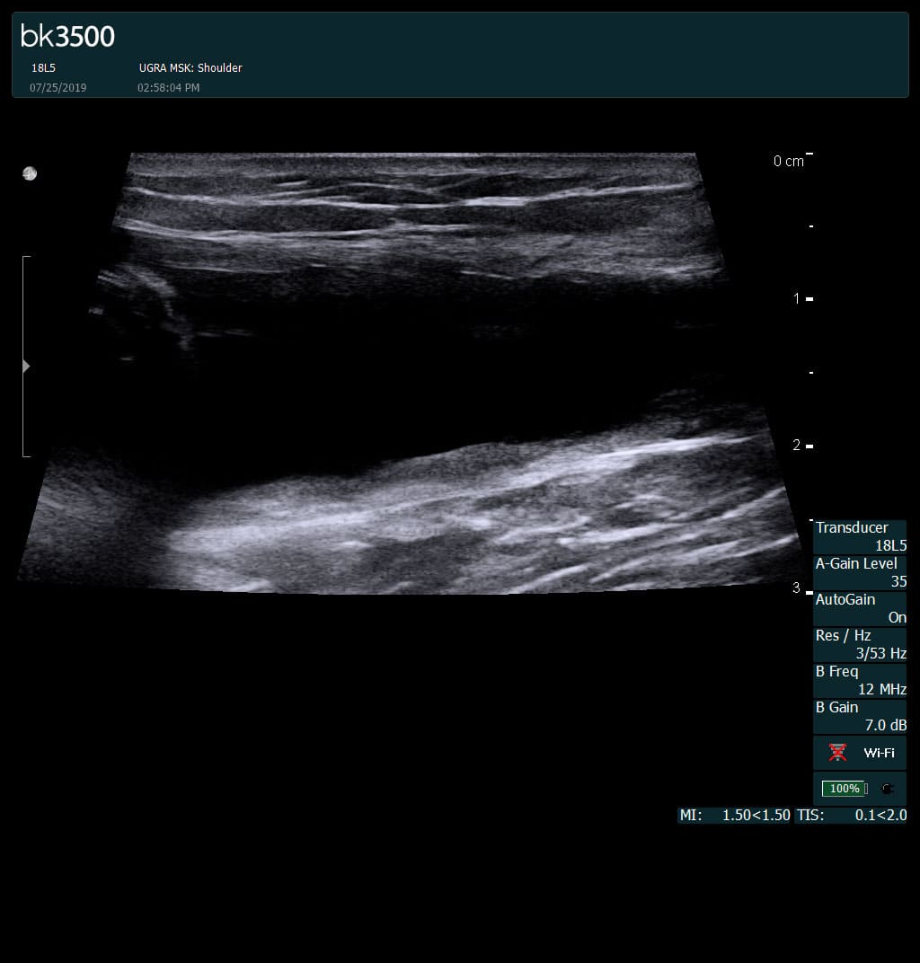
Separation of muscle ends due to tear elicited
on dynamic sonography examination


 Request Telehealth