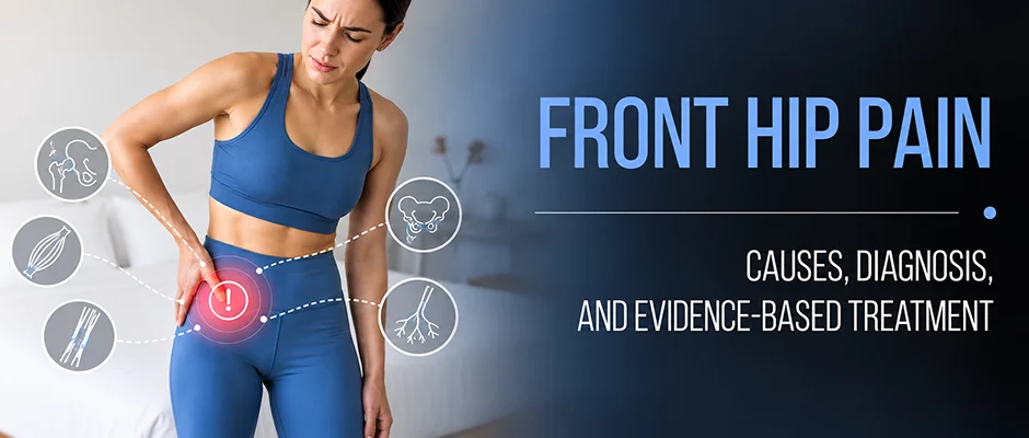
Pain at the front of the hip, in the crease where your thigh meets your pelvis, can come from muscle, tendon, joint, bone, or nerve, and the right treatment depends entirely on which one. This guide walks through the common and easily-missed causes, how they are told apart, and what actually helps.

Anterior hip pain is pain in the front of the hip, in the crease where the thigh meets the pelvis or deep in the groin. The terms are used interchangeably in this guide. It may be a mild, persistent background pain or it may be a more intense pain that interferes with your activities and halts your stride.
At nydnrehab, it’s one of the most common and most frequently mismanaged conditions we treat. That’s because the term “front hip pain” is not a diagnosis, it’s a location. The key to effective treatment is pinpointing the specific structure that is causing the problem.
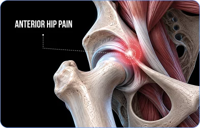
Muscles, tendons, bursae, the hip joint, surrounding nerves, the lumbar spine or even referred pain from an internal organ can all cause pain in this area. In some patients, it is not even a visible structural lesion, but rather the abnormal movement of the joint, the loading of the capsule or the irritation of the nerves that conventional imaging methods may not detect. Correct diagnosis is the key.
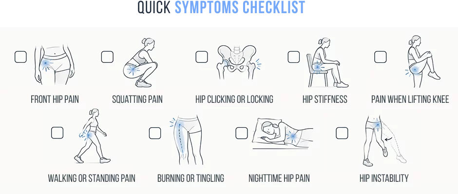
If pain does not improve with changing position, fever is present with hip pain, unable to bear weight or symptoms worsen at rest, this may be a sign of an underlying problem such as stress fracture, avascular necrosis, septic arthritis or inflammatory disease. These warning signs are not listed in full elsewhere in this article. If you have any of these, please don’t wait to be evaluated.
The following are the most common causes of anterior hip pain in the clinical setting. Every entry details the nature of the condition, its causes, its symptoms and the treatment. See the Differential Diagnosis table below for a rapid overview of pain location, type and probable diagnosis
Hip Flexor Strain and Tendinopathy
The hip flexors are mostly the iliopsoas and rectus femoris. They are active in running, cycling, kicking and climbing the stairs and if they are asked to do more than they can comfortably do, microtears occur in the muscle fibre or tendon. This causes pain when lifting the knee, climbing the stairs or getting up from a chair.
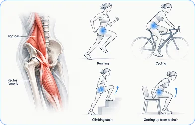
Many patients are not aware that iliopsoas tendinopathy can occur in parallel with other injuries. The iliopsoas tendon lies directly over the anterior hip capsule and tendon symptoms can develop as a result of intra-articular pathology or hip microinstability which causes the surrounding musculature to overload and attempt to dynamically stabilise the hip. If iliopsoas symptoms are out of proportion or do not improve with traditional tendon loading, then this is a consideration.
Common presentation: localised anterior hip discomfort, aggravated by resisted hip flexion; tenderness on palpation of the tendon; stiffness after rest and movement.
What sets it apart: pain can be reliably reproduced with resisted hip flexion and is not associated with clicking or locking often. Labral tears do not give the sensation of instability, but can occur in combination with labral tears.
Recovery: 4-8 weeks of load management and targeted rehabilitation. Chronic tendinopathy usually needs a longer more structured loading programme.
A Mechanical Driver that’s Easy to Miss: Hip Microinstability
Hip microinstability is defined as excessive translation of the femoral head in the acetabulum, which is not dislocating but is beyond the passive and active limits of the hip joint’s stabilisers. It is one of the less talked about causes of chronic anterior hip pain and is especially important in the patient whose MRI scans seem surprisingly benign for the amount of pain that they are suffering.
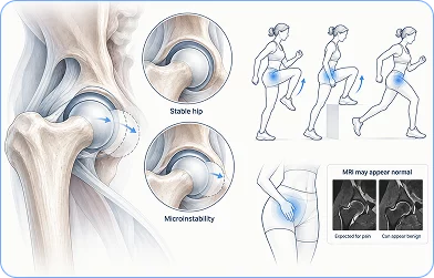
Here’s why it causes pain. The abnormal translation that is repetitive places extra mechanical load on the anterior capsule, the iliofemoral ligament, the labrum and the chondrolabral junction, especially during extension and external rotation. This results in capsular strain, overload of the labrum, synovial irritation and secondary iliopsoas overactivity, as other muscles attempt to compensate for the lack of passive stabilisers.
This is clinically significant, but not always understood, due to the anatomy of the anterior capsule. The area is well innervated with articular branches of the femoral and obturator nerves. Repetitive capsular loading is then a direct source of nociceptive signalling and can cause real anterior hip and groin pain in the absence of significant imaging findings of a structural tear. This is the story of so many frustrating cases, where there is persistent disabling hip pain, an MRI that is relatively unremarkable, and months of treatment that has failed, because it was treating the wrong structure.
Microinstability should be considered as a primary or contributing cause when pain persists after proper rehabilitation of the labrum or iliopsoas or cartilage. In these cases treatment should focus on neuromuscular stabilisation of the hip joint, rather than passive treatment of the underlying mechanical problem.
Femoral Articular Branches: A Neural Source That Imaging Won’t Show
A key question that follows the idea of microinstability is how does repeated loading of the capsule actually lead to pain? The innervation of the anterior hip capsule is a part of the answer.
Articular branches from the femoral, obturator and accessory obturator nerves are the main nerves of the anterior capsule. The femoral articular branches provide special innervation to the anterosuperior capsule, which is rich in nociceptive receptors and is often a site of painful intra-articular hip disorders. These small sensory branches pass through the deep iliopsoas area and then into the capsule, thus establishing an anatomical connection between capsular pathology, the iliopsoas fascial interface and the anterior hip pain the patient is feeling.
Importantly, the irritation of these articular branches is not a true femoral neuropathy. The features that suggest nerve root compression or peripheral neuropathy of the nerve are deep anterior hip or groin pain, weakness of the quadriceps, thigh sensory loss, and change in the patellar reflex; however, these are absent in a patient. This is very significant for diagnosis.
Identification of the neural contribution to anterior capsular pain can also be a useful addition to the clinical picture in patients in whom symptoms cannot be explained by traditional imaging, and provides a new therapeutic option that would otherwise be completely overlooked by structure-based therapy.
Acetabular Labral Tear
The labrum deepens the hip socket and is also a suction seal, helping a lot with hip stability. A sudden trauma, repeated rotational forces or bony abnormalities like FAI are the causes of tears. They may also form insidiously in the background of a hip microinstability, which is an abnormal loading of the labral tissue with repetitive stress over time, and is an easy-to-miss diagnosis if imaging is used as a standalone assessment.
Common symptoms: deep groin pain; clicking, catching or locking; pain when sitting for long periods or pivoting; feeling the hip is unstable.
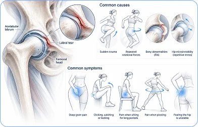
Diagnosis: MR arthrography (MRA) is the gold standard.
Conservative treatment: the cornerstone of conservative treatment is physiotherapy to address hip stability, neuromuscular control, and movement retraining. Hip arthroscopy is considered when conservative treatment has been successful for 3-6 months but it has not helped.
Femoroacetabular Impingement (FAI)
FAI is when the ball and socket come into abnormal contact during movement because of a bony abnormality. Cam impingement is an irregular (abnormal) hip head, pincer impingement is when there is too much bone that extends over the rim of the acetabulum and many patients have both, combined impingement.
Typical presentation: deep pinching pain with hip flexion and internal rotation; pain on sitting for long periods, squatting or tying their laces; gradual onset over months, not from a single identifiable injury.
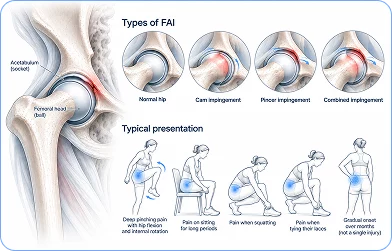
Important: FAI morphology is seen in many people who are not symptomatic. The clinical history, physical examination and response to treatment are all important in making a diagnosis, and a positive MRI is not enough to make the diagnosis.
Treatment: NICE and APTA guidelines recommend first line treatment to be physiotherapy. Surgery is only considered after 3 months or more of well directed and structured rehabilitation has failed to achieve sufficient improvement.
Hip Osteoarthritis
Osteoarthritis is a progressive wearing down of the cartilage in the hip joint. It most often occurs after the age of 50, but it also can occur at a younger age after major trauma or in the setting of longstanding FAI which has gradually worn down the joint surface.
Common symptoms: pain in deep groin, morning stiffness (gone in 30 minutes), loss of internal rotation, pain with weight bearing, and problems with daily activities like getting on and off shoes or socks.
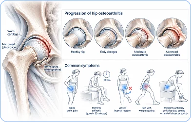
Management: all major clinical guidelines (OARSI, NICE, APTA) strongly support exercise therapy and weight management. Although physiotherapy cannot stop cartilage deterioration, it is able to effectively relieve pain and enhance function, and sometimes even prevent the necessity of surgery. For severe OA that does not improve with conservative treatment, total hip replacement is one of the most successful surgical options available.
Iliopsoas Bursitis and Snapping Hip Syndrome
The iliopsoas bursa, the largest bursa in the human body, is located directly below the iliopsoas tendon at the anterior (front) hip. If it becomes inflamed, it causes a deep anterior hip pain, which may be hard to differentiate from tendon or joint pain. Many patients also experience a characteristic audible or palpable “snapping hip” (coxa saltans interna) as the iliopsoas tendon snaps over the iliopectineal eminence when the hip moves.
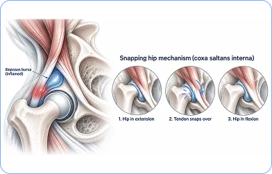
It’s important to note that like tendinopathy, bursitis doesn’t always occur alone. Both conditions can result from intra-articular pathology or microinstability and treating the bursa alone will not alleviate symptoms if it is caused by a deeper structural issue.
Typical presentation: deep groin pain, snapping when flexing and extending the hip, anterior hip pain, and aggravated by repetitive flexion (running, climbing up and down stairs).
Treatment: activity modification, physiotherapy focusing on hip stability and movement mechanics, and if conservative treatment fails, ultrasound guided corticosteroid injection can help provide a short-term solution.
Stress Fracture of the Femoral Neck
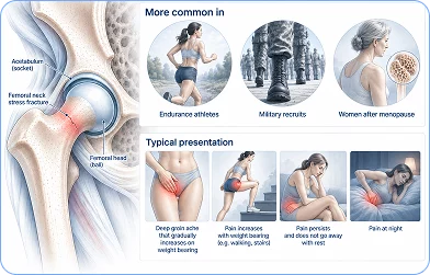
Stress fractures of the femoral neck occur when the bones are overloaded with repeated stress which is greater than the body’s remodelling process. Their appearance is most commonly seen in endurance athletes, military recruits and women after their menopause, and they are severe enough to be considered a category for concern.
Presentation: deep groin ache that gradually increases on weight bearing, which does not go away with rest, pain at night, localised tenderness at the femoral neck.

Meralgia Paraesthetica
Meralgia paraesthetica is caused by compression of the lateral femoral cutaneous nerve (LFCN) as it passes under the inguinal ligament, near the anterior superior iliac spine (ASIS). It does not cause joint or tendon pain like most of the other conditions in this section, but causes a unique sensory disturbance on the outer thigh, which is usually diagnostic when it is suspected.
The typical presentation is of burning, tingling or numbness on the outside of the thigh, a patch-like area of altered sensation with a fairly consistent distribution, worsening with prolonged standing or walking, and no muscle weakness.
Common causes: tight belts or waistbands, pregnancy, quick weight gain and prolonged hip flexed positions at work or in the car.
Treatment: first, the source of mechanical compression needs to be identified and treated. Nerve mobility exercises and activity modification usually form the basis of conservative management. The majority of cases are treated without surgery.
Sports Hernia (Athletic Pubalgia)
Although it’s called a sports hernia, it’s not a true hernia. A strain or partial tear of the muscles and tendons that attach to the pubic symphysis, usually the lower abdominal wall or adductor musculature. It forms when there is repetitive shear loading across the pelvis, and is most prevalent in athletes whose sport requires kicking, sprinting or quick changes of direction.
Typical presentation: groin and lower abdominal pain, which increases with kicking, sprinting or abdominal exercises, tenderness over the pubic symphysis, symptoms improve with rest but return rapidly when returning to sport.
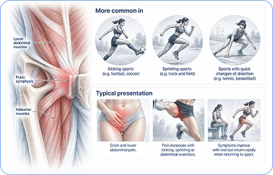
Treatment: progressive core and adductor rehab is primary treatment.
Less Common but Important Pain Generators
When the more common diagnoses have been excluded, or when a patient’s presentation doesn’t quite fit the expected pattern, the following sources deserve consideration.
Iliohypogastric Nerve Entrapment
The iliohypogastric nerve, arising from T12 to L1, predominantly L1, does not innervate the hip joint itself, but it can produce pain that patients clearly perceive as anterior hip or pelvic discomfort. After emerging from the lateral border of the psoas, it crosses the quadratus lumborum and travels between the abdominal wall muscle layers, giving off a lateral cutaneous branch to the superolateral gluteal and iliac crest region and an anterior branch to the suprapubic area.
When this nerve is irritated or entrapped typically near the iliac crest or within the abdominal wall fascial planes the resulting pain can be easily mistaken for a hip or groin problem. The key distinguishing features are a superficial, burning, or radiating quality, and the ability to reproduce symptoms by palpating along the nerve’s anatomical course. When imaging provides no satisfying explanation for a patient’s anterior hip symptoms, this neural source is worth considering.
Lumbar Spine Referral
The lumbar spine is a more frequent mimicker of hip pathology than many patients expect. Upper lumbar nerve root irritation particularly at L1 through L3 can produce pain in the groin, anterior hip, or anterior thigh without generating any back pain at all. Lumbar disc and facet-related pain can refer in a similar distribution without producing a true radiculopathy.
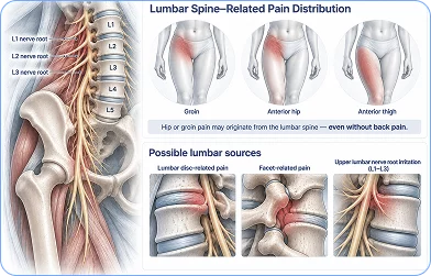
A spinal origin should be considered whenever anterior hip pain is accompanied by back symptoms, neurologic findings, or pain that changes with lumbar movement. It is worth noting that S1 radiculopathy typically produces posterior rather than anterior symptoms, a useful differentiating feature.
Sacroiliac Joint Referral
The sacroiliac joint is not a common cause of anterior hip pain, but it is an important one to keep in mind. While SI joint symptoms most often present in the posterior pelvis and buttock, a subset of patients experience referral patterns that extend toward the groin or anterior hip, an overlap that can make SI joint dysfunction genuinely difficult to distinguish from intra-articular hip pathology on the basis of symptoms alone. Examination of the SI joint and lumbopelvic region is therefore appropriate when anterior hip pain cannot be fully explained by local hip findings particularly when posterior pelvic or buttock pain accompanies the presentation.
Other Serious Causes to Rule Out
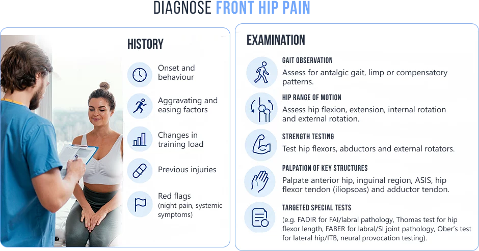
Diagnosis begins with clinical assessment, not imaging.
Imaging, ordered when clinically indicated:

If you have anterior hip pain without red flags that is mild to moderate, the following are reasonable measures to take until you are seen by a professional or until you have begun to recover.





Phase 1 Gentle Activation (Acute Stage)
The focus during this stage is on keeping the blood flowing, moving joints gently in their range of motion, and getting muscles moving as early as possible, while avoiding adding weight to the irritated structures.
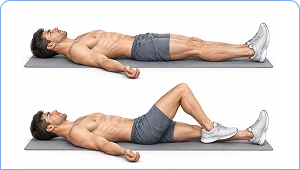

How: on your back, move one heel towards your buttock and bend the knee and hip slowly, then extend.
Dosage: 10-15 repetitions per set
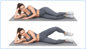
How: with knees bent and feet on top of each other, lie on your side. With feet together, raise the upper knee towards the ceiling. Hold for 2 seconds and slowly lower.
Dosage: 3 sets of 15 reps
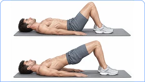
How: lie on back with knees bent and feet flat. Press feet together into heels and lift hips off the floor. Lower with control, hold for 3-5 seconds.
Dosage: 3 sets of 12 reps
Phase 2 Strength and Stability (Improving Stage)
After pain has subsided, and movement is more comfortable, strengthening exercises begin for the muscles that support and protect the hip joint.
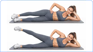
How: lying on your side with your body in a straight line, lift top leg to around 30-40 degrees. Hold for 2 seconds, slowly lower.
Dosage: 3 sets of 15 reps
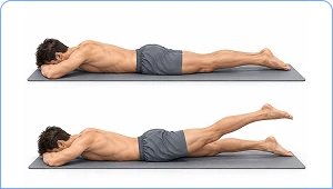
How: lying face down, straighten the legs and squeeze the glute muscles of one side and raise that leg off the ground a few cm. Hold for 3 seconds, slowly lower.
Dosage: 3 sets of 12 reps
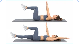
How: on your back with arms up to the ceiling, hips and knees bent at 90 degrees, slowly lower one arm over your head while extending the other leg towards the floor, keeping the lower back flat.
Dosage: 3 x 8-10 reps on each arm
A common mistake is to over-stretch the hip flexors when experiencing anterior hip pain. Tendons and/or anterior capsule are commonly already irritated and stretching them often aggravates the situation. Strengthening and load management are the first, stretching is the second (if applicable).
Phase 3 Functional Loading (Return to Activity)
This is a transition period between strengthening and re-entering the demands of sport, work or higher level activity.
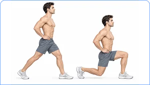
How: stand in a staggered position with one foot in front and one behind. Straighten back knee toward floor, keeping front shin straight. Push back up through the front heel.
Dosage: 3 sets of 10 on each side
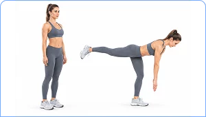
How: stand on one foot and bend forward from the hip with the other leg behind you to balance. Slowly lower into a stretch in the standing leg’s hamstring and slowly return to standing
Dosage: 3 sets 8-10 reps on each side
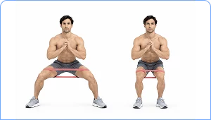
How: place a resistance band just above the knees. Feet should be shoulder-width apart with knees slightly bent. Take 10-15 steps side steps, leading foot first, following foot second.
Dosage: 3 sets іn each direction
When to stop and seek advice: If pain worsens during or after exercise, and settles within 24 hours or if new symptoms occur, e.g. swelling, giving way or changed sensation, stop programme and seek advice from a clinician before continuing.
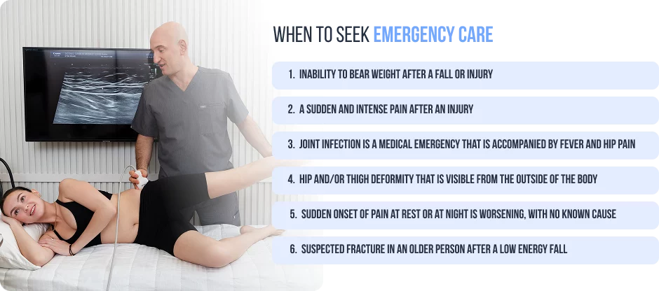
Most cases of front hip pain are not a medical emergency. Yet there are some presentations that must be evaluated the same day or immediately or else there can be serious consequences.
If you have any of the following symptoms, get emergency medical help right away:
First, we determine the precise cause at nydnrehab, as treatment is always underwhelming when not tailored to a specific diagnosis. So many patients come in on a protocol which has been followed for months and months which do not match what is going on in their hip.
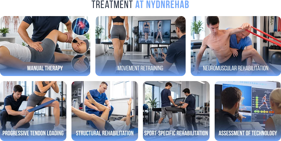
We do more than the typical orthopaedic evaluation. We will look at your movement, the way you load the hip when performing functional tasks, and where your movement system is compensating. If microinstability, neural irritation or referred pain from the spine or SI joint is suspected, we expand our assessment to include lumbopelvic examination, neural provocation testing and dynamic ultrasound evaluation, which are not possible with static MRI.
Treatment at nydnrehab could involve:
Most hip problems on the front of the hip are treated with physiotherapy as the basis of treatment. However, for some, medical treatment can be an adjunct to surgery, especially when the pain is very intense, when treatment is not advancing as quickly as it should, or when the diagnosis of the underlying problem would suggest it.
Medications:
Injections:
On PRP: evidence is promising but not yet definitive. At nydnrehab, we discuss the current evidence openly with every patient considering this option.
Injections are most useful as a short-term pain bridge to allow for rehabilitation and are not a treatment in and of themselves.
Surgery is indicated for structural problems that have failed to respond to 3–6 months of proper conservative treatment:
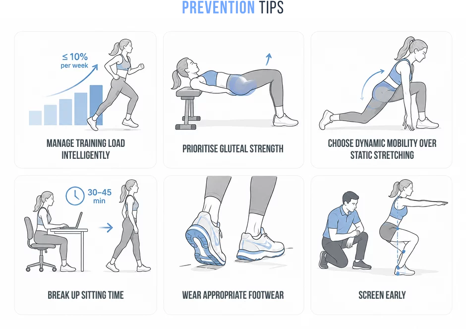
Prevention is most effective when it’s specific. The methods used by a competitive runner are not necessarily applicable to a desk worker or an older person re-entering activity after a period of inactivity. Though there are some general rules:
Yes and, more frequently than many patients would think. L2 and L3 nerve roots in the spine provide sensation to the front of the hip and groin, and so a problem with the disc or nerve root at these levels can cause pain in the front of the hip, without any pain in the back at all. The sacroiliac joint may also cause pain to the front of the hip, especially if the pain is also felt in the back of the pelvis or buttocks. If the clinical picture is not clear, examination of both the lumbar spine and SI joint is an important component of a comprehensive hip examination.
Yes. Anterior hip pain can be caused by several neural structures, but not identified on imaging. Meralgia paraesthetica is caused by the lateral femoral cutaneous nerve, which causes burning and numbness on the outside of the thigh. When the iliohypogastric nerve is trapped near the iliac crest or in the abdominal wall, it may cause pain in the front of the hip and abdomen. Small sensory nerves (and femoral articular branches) that innervate the anterior capsule can be a source of deep groin pain in the setting of an otherwise normal MRI. Anterior hip pain is often attributed to a different source than the neural origin and needs to be specifically tested for.
A labral tear can’t be diagnosed just from symptoms. Deep groin pain, clicking, catching or a feeling of instability are suggestive, but these can also be seen in FAI, osteoarthritis and microinstability. MR arthrography (MRA) is the most accurate imaging investigation available. Diagnosis requires correlation between imaging findings, clinical history, and physical examination.
Hip microinstability refers to excessive motion of the femoral head in the socket, but not enough to cause a dislocation, that is enough to repeatedly stress the anterior capsule, labrum and surrounding structures. It is a largely overlooked source of chronic anterior hip pain especially when the MRI is relatively normal. Microinstability can be a factor if the pain has not been resolved with treatment aimed at the labrum, iliopsoas or cartilage. Specific clinical assessment and a rehabilitation strategy involving neuromuscular joint stabilisation instead of passive treatments are required.
Not always. Passive stretching of the hip flexors is often advised for anterior hip pain, but not always. Aggressive stretching may aggravate symptoms if you have an active iliopsoas tendinopathy, anterior capsular irritation or a labral tear. Strengthening and load management are typically more suitable first line interventions. If you experience pain that worsens with regular stretching, discontinue and get medical advice.
Depending on the cause. Gentle walking is good and should be continued for most muscular and tendon conditions. Walking may worsen symptoms of stress fractures and active bursitis. If walking regularly aggravates the pain and it does not resolve within an hour of stopping, it is a good idea to have it assessed professionally instead of persisting in walking.
In most cases, yes. Swimming, bicycling (with proper seat height adjustment), elliptical training are generally tolerated by low impact activities. Depending on the diagnosis, temporarily modifying high impact activities, deep squatting, and heavy hip flexion loading may be necessary. Rest is never the answer, as long as you select the right activities for your situation.
Recovery timelines vary considerably depending on the diagnosis, severity, and how early treatment begins.
These are general estimates. Individual recovery depends on diagnosis accuracy, treatment quality, compliance with rehabilitation, and the presence of any contributing factors such as microinstability or referred pain.
Yes. Prolonged sitting maintains the hip in flexion and shortens the iliopsoas. Over time, this contributes to hip flexor tendinopathy, anterior impingement, and bursitis. People with FAI or anterior capsular sensitivity are particularly prone to pain with prolonged sitting due to the compression of the anterior joint structures. Regular movement breaks, standing briefly every 30–45 minutes, are one of the simplest and most effective preventive measures available.
Sleeping on your back with a pillow under the knees reduces anterior hip tension. If you prefer side-lying, place a pillow between your knees to prevent the hip from dropping into adduction. Avoid sleeping on your stomach, this places the hip in sustained extension and internal rotation, which can aggravate several anterior hip conditions including iliopsoas tendinopathy, labral tears, and FAI.
Surgery becomes appropriate when a clearly identified structural problem, such as a labral tear, cam deformity, or advanced osteoarthritis, has not responded to at least 3–6 months of well-executed conservative care. The decision should always be made collaboratively between you, your physiotherapist, and your orthopaedic surgeon, based on your symptoms, imaging findings, functional limitations, and personal goals. Surgery is rarely the first option and should never be pursued before a genuine trial of structured rehabilitation.
If your front hip pain has persisted for more than two weeks, is affecting your daily activities, or simply isn’t responding to self-care, a professional assessment is the logical next step.
At nydnrehab, our clinical team specialises in identifying the precise source of anterior hip pain and building treatment plans that are matched to your diagnosis, not a generic protocol. We combine detailed biomechanical assessment, hands-on treatment, and evidence-based rehabilitation to help you return to full function as efficiently as possible.
Whether your pain has a clear structural cause or has been frustratingly unexplained by imaging, we have the clinical tools and diagnostic depth to find the answer and act on it.
Contact nydnrehab today to schedule your assessment and take the first step toward understanding and resolving your hip pain.
Request an appointment»

Our location: 11 West 25th Street 5th floor, New York, NY 10010
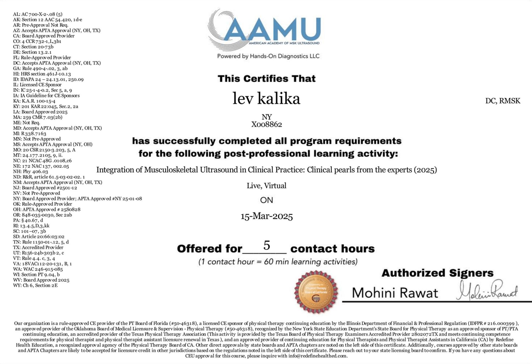
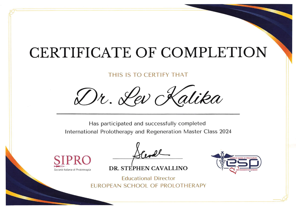
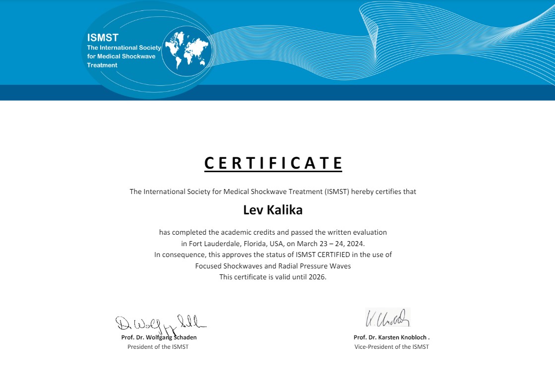
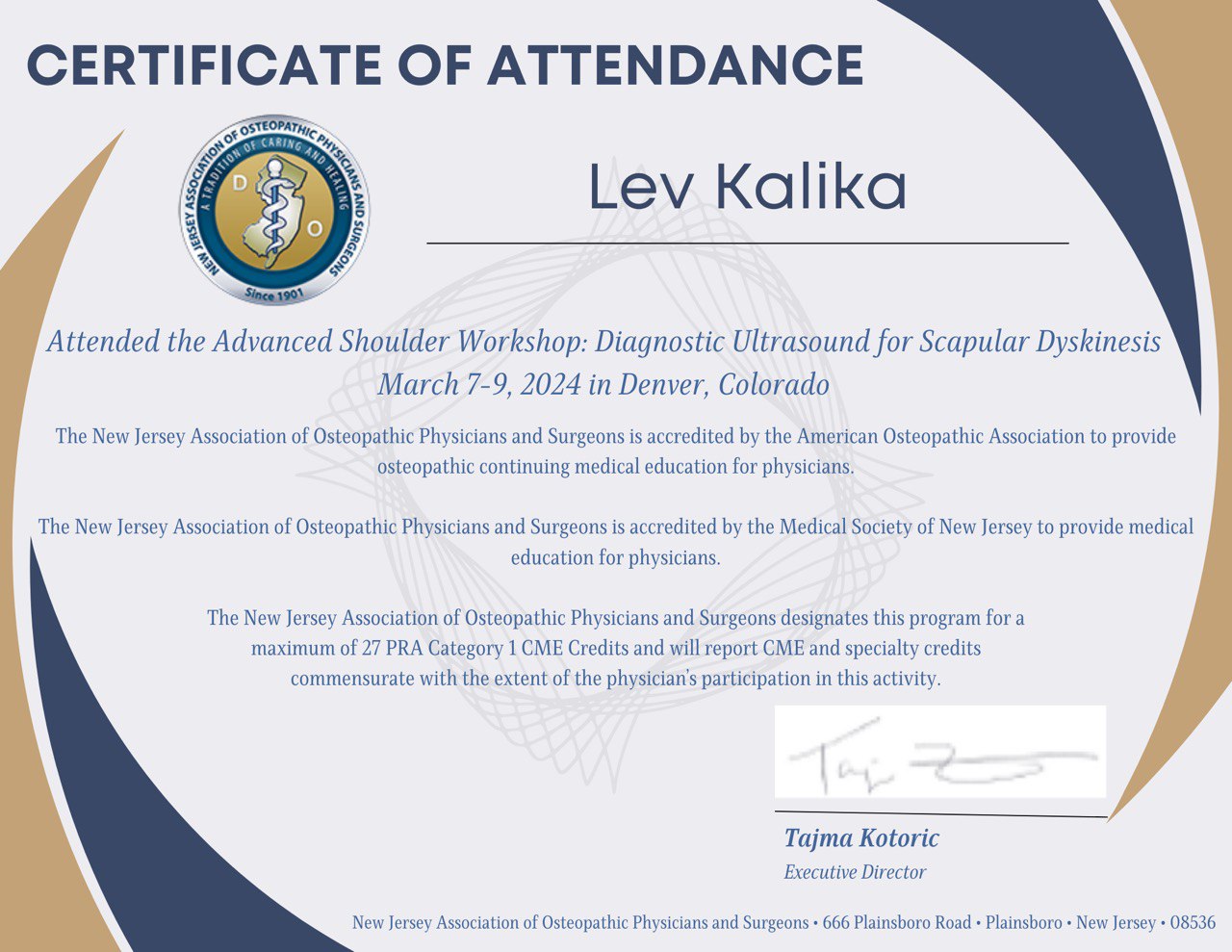
Dr. Lev Kalika is a world-recognized expert in musculoskeletal medicine. with 20+ years of clinical experience in diagnostic musculoskeletal ultrasonography, rehabilitative sports medicine and conservative orthopedics. In addition to operating his clinical practice in Manhattan, he regularly publishes peer-reviewed research on ultrasound-guided therapies and procedures. He serves as a peer reviewer for Springer Nature.
Dr. Kalika is an esteemed member of multiple professional organizations, including:

Below is a prime example of how ultrasound can take the guesswork out of diagnosis.

A bad physical therapy experience is one of the primary causes of unnecessary surgery

In this instance, an athlete was originally diagnosed with minor quadriceps muscle strain and was treated for four weeks, with unsatisfactory results. When he came to our clinic, the muscle was not healing, and the patients’ muscle tissue had already begun to atrophy.
Upon examination using MSUS, we discovered that he had a full muscle thickness tear that had been overlooked by his previous provider. To mitigate damage and promote healing, surgery should have been performed immediately after the injury occurred. Because of misdiagnosis and inappropriate treatment, the patient now has permanent damage that cannot be corrected.
The most important advantage of Ultrasound over MRI imaging is its ability to zero in on the symptomatic region and obtain imaging, with active participation and feedback from the patient. Using dynamic MSUS, we can see what happens when patients contract their muscles, something that cannot be done with MRI. From a diagnostic perspective, this interaction is invaluable.
Dynamic ultrasonography examination demonstrating
the full thickness tear and already occurring muscle atrophy
due to misdiagnosis and not referring the patient
to proper diagnostic workup
Demonstration of how very small muscle defect is made and revealed
to be a complete tear with muscle contraction
under diagnostic sonography (not possible with MRI)
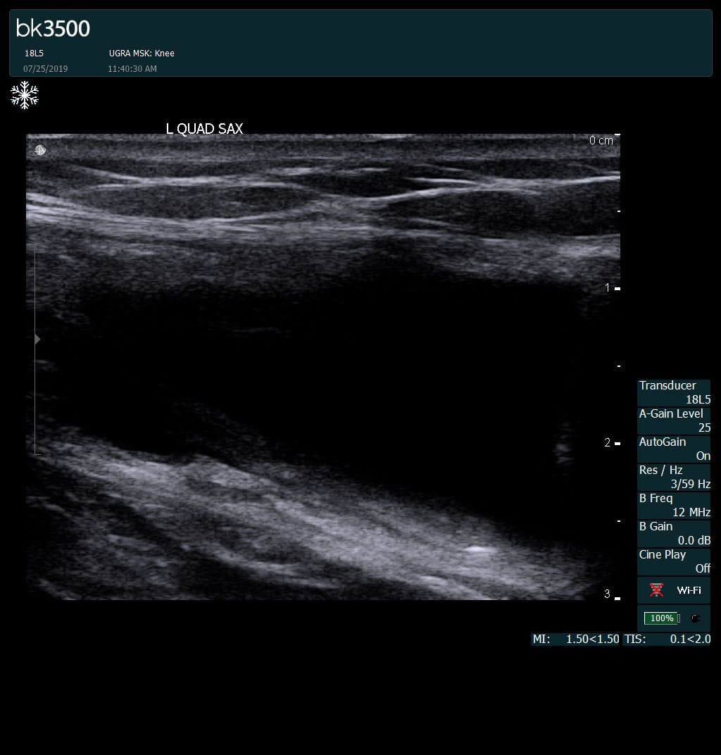
Complete tear of rectus femoris
with large hematoma (blood)
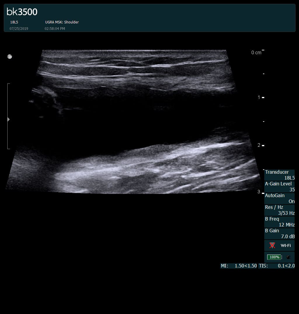
Separation of muscle ends due to tear elicited
on dynamic sonography examination

 Request Telehealth