Trigger points are small but they can generate a lot of trouble. A taut band in the upper trapezius, no bigger than a grape, can produce headaches, shoulder restriction, and referred pain down the arm that mimics a rotator cuff tear. Identifying that band accurately, and reaching it safely with a needle, is harder than it sounds – and the difference between accurate and approximate matters considerably when the target sits near the brachial plexus or the apex of the lung.
This article explains the science behind trigger points, what ultrasound guidance actually adds to dry needling, and how NYDNRehab integrates these tools into a broader treatment approach.
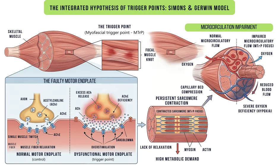
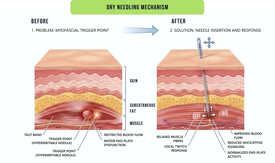
The most important theory on trigger points is the integrated model of David Simons and later Gerwin (2023). The core of that model is a faulty motor endplate that secretes excess acetylcholine. Too much acetylcholine causes the sarcomeres to stay contracted and a small group of muscle fibres is unable to relax – the compression of the capillary bed at the same time reduces the delivery of oxygen exactly when it’s needed. The muscle is unable to repolarize completely.
The reason why trigger points are self perpetuating is because of calcium dysregulation. Following a normal contraction, the calcium pump in the sarcoplasmic reticulum removes calcium from the cytoplasm, using ATP. In an ischemic zone (contracted zone), ATP is depleted, and the pump stops. Calcium stays elevated. The sarcomere remains contracted. In this condition, a patient may complain of a constant aching pain at the base of the skull, and may not be able to rotate the neck at all, even on days when they haven’t done anything rigorous – the low level, always present tension is the self sustaining cycle made real.
Pressure sensitivity is the sensation the patient reports, which is stiffness or achiness at rest, and often referred pain that is not dermatomal in distribution. The non-dermatomal referral is the reason that trigger points are misdiagnosed as radiculopathy or nerve entrapment with some frequency. Reviewing the most prominent theories, Zhai et al. (2024) found that none of the integrated hypothesis, central sensitization model, or energy crisis variants are able to account for all the phenomena observed. The clinical implication is that trigger points are likely to be a combination of peripheral and central mechanisms and not a purely local tissue problem.
The treatment of a trigger point without the understanding of the cause is similar to fixing a tire without removing the nail. Trigger points are most likely to recur due to:
The thoracic spine is stiff and the cervical paraspinal muscles and suboccipital muscles must compensate during rotation, which is called altered joint mechanics. Those muscles do not develop trigger points due to weakness, but rather due to overloading caused by a lack of mobility elsewhere.
Fascial restriction: when fascia is dense or adherent, glide is restricted and mechanical stress is focussed at particular points within a muscle. A trigger point is where the load is greatest.
Scapular dyskinesis: if the serratus anterior and lower trapezius do not stabilize the scapula during shoulder elevation, then the upper trapezius and levator scapulae will do so. One of the most common trigger point generators encountered in clinical practice is chronic overuse of those muscles.
Gait dysfunction: tibialis posterior weakness, hip abductor inhibition or leg-length discrepancies alter the distribution of load through the lower kinetic chain. Piriformis, gluteus medius and hamstring trigger points often can be traced to gait asymmetry and not local injury.
Central sensitization reduces the threshold for a motor endplate to become dysfunctional (psychosocial load and disruption of sleep). Patients who are poorly managed with stress or sleep deprivation can develop trigger points more quickly and more easily, and are less likely to resolve the trigger.
Any treatment plan that doesn’t address these upstream drivers will bring temporary relief and frustration to the patient and the clinician.

Palpation-based dry needling is a standard technique that requires the clinician to find a taut band, estimate depth based on surface anatomy, and insert the needle without real-time knowledge of the needle’s trajectory. This is fairly satisfactory in superficial, clearly defined muscles where there are no important structures in close proximity. It is less effective in other locations.
Dynamic (not static pre-procedure) musculoskeletal ultrasound demonstrates:




“Palpation-based” is not synonymous with the lack of skill. Traditional landmarks are competent in appropriate anatomical territories by experienced clinicians. The comparison is not between the level of training of the practitioner, but rather between what each approach can and cannot confirm.
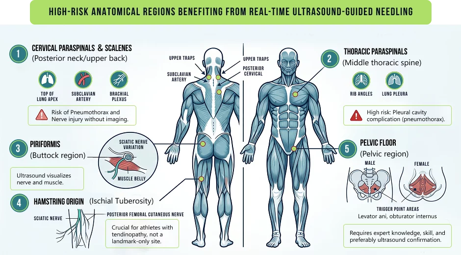
Some areas are sufficiently close to vulnerable structures where the margin for error is large and meaningful when imaging in real time is not used.
The local twitch response is the sensation most closely linked to dry needling and is characterized by a small, involuntary muscle twitch that occurs when the needle is inserted into the taut band. Patients have reported it as a cramping, jumping or grabbing feeling. The first time it can be a surprise. It is not a bad sign, there is some evidence that it is related to better clinical outcomes, although the relationship is not a linear one.
Referred pain reproduction occurs frequently. Many patients report the familiar pain as it radiates down to the temple or behind the eye as a result of the accurate needle placement of a trapezius trigger point – the same pattern that led them to the clinic. This indicates that the needles are in place and helps to reassure patients once they have understood the significance of the procedure.
The first 24-48 hours post-treatment can include delayed onset muscle soreness that is comparable to exercise-induced delayed onset muscle soreness. Immediate relief is experienced by some patients, while others experience improvement within 2-3 days. It is common to experience fatigue on the day of the treatment and patients should be advised to avoid strenuous activity for the rest of the day. By the next day, most people are back to normal.




Dry needling should not be the initial treatment for all patients and the presence of trigger points does not indicate that needling is the next step. A detailed movement assessment and biomechanical evaluation frequently alters the treatment plan.
Dry needling is best used as part of a treatment episode and not as an isolated treatment. The typical treatment at NYDNRehab is the combination of needling with:
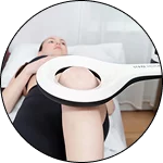
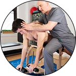
Palpation is the most common technique used for dry needling. The clinician uses his or her hands to feel for the taut band, estimates the depth, introduces the needle, and checks the location by listening to the patient and feeling the needle. This is good enough in simple anatomical areas where there is skilled experience.
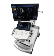
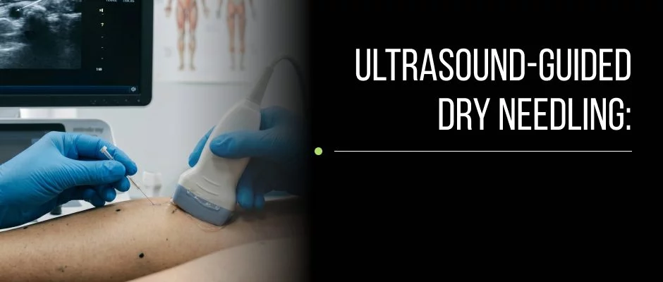
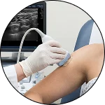
NYDNRehab incorporates real time dynamic musculoskeletal ultrasound as a part of each needling treatment. This isn’t a marketing difference, it’s a procedural one. The clinician observes the tip of the needle during insertion, verifies the location of the needle within the target area and modifies the trajectory if adjacent structures (nerves, vessels, fascial layers) are closer than what surface anatomy would suggest. The imaging also documents the response of the tissue during the treatment – changes in the echogenicity of muscles, movement of the fascia, local hyperemia on Doppler – which are not palpable.
The clinic’s overall approach is focused and not about quantity. Less, more focused interventions, based on objective imaging and movement data, tend to be more lasting than repeated generalized interventions. This attitude is carried over into patient selection, treatment planning, and when to combine platelet-rich plasma or prolotherapy with needling to achieve a better result.
Request an appointment»

Our location: 11 West 25th Street 5th floor, New York, NY 10010
The tools are similar, but the theory and target tissues are different. Dry needling is a technique that is based on anatomical and neuromuscular principles and uses anatomical and neurological landmarks to target myofascial trigger points and tendinous structures. Acupuncture works through a traditional Chinese medicine system with the use of meridian points. The overlap is in the actual act of putting the fine needle in; it’s not in the clinical rationale and point selection.
The needle is of the same gauge as with normal dry needling. The ultrasound imaging is performed with a hand-held probe placed on the surface of the skin, and is not painful beyond the pressure of the probe. Imaging is added to the procedure, making it not more painful.
This depends a lot on the duration of the trigger points, the root cause of the trigger points and if there is co-pathology (tendinopathy, fascial restriction) being treated. Acute presentations with a definite mechanical cause tend to respond in 2-4 sessions. If chronic, multi-site myofascial pain with central sensitization are present, then a longer treatment episode may be needed along with rehabilitation.
This will be sore and normal after treatment. If the pain is increasing, there are new neurological symptoms or the pain is still present after 72 hours, this is considered genuine worsening and should be reported to the treating clinician. Dry needling is a safe procedure when performed by a trained clinician with the necessary precautions; in high-risk anatomical areas, ultrasound guidance further decreases risk of adverse events.
A trigger point injection is a procedure in which a substance (usually local anesthetic, sometimes corticosteroid or saline) is pushed into the needle. The mechanical effect of the needle on the taut band is the main mechanism of dry needling and there is no injectate used. Both can cause a twitch reaction and referred pain reproduction during the procedure.
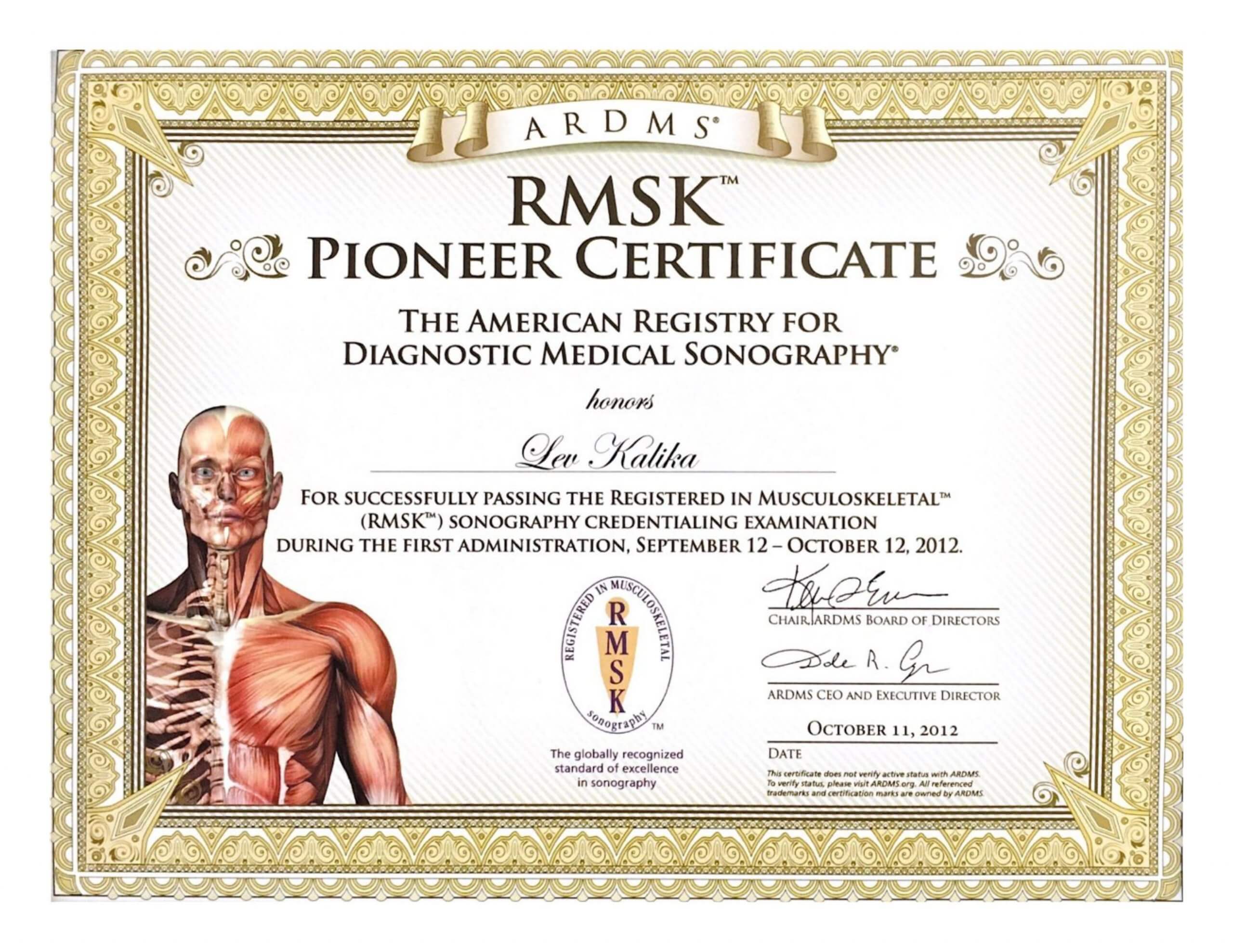
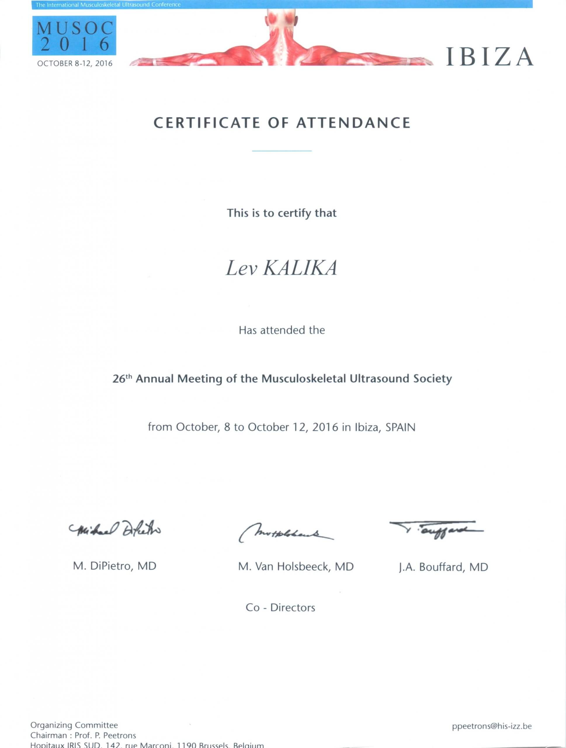
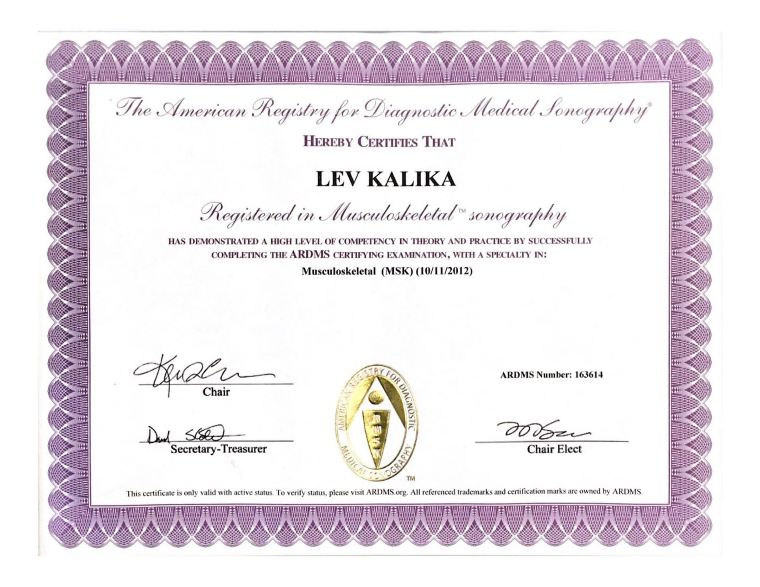
Dr. Lev Kalika is a world-recognized expert in musculoskeletal medicine. with 20+ years of clinical experience in diagnostic musculoskeletal ultrasonography, rehabilitative sports medicine and conservative orthopedics. In addition to operating his clinical practice in Manhattan, he regularly publishes peer-reviewed research on ultrasound-guided therapies and procedures. He serves as a peer reviewer for Springer Nature.
Dr. Kalika is an esteemed member of multiple professional organizations, including:

Below is a prime example of how ultrasound can take the guesswork out of diagnosis.

A bad physical therapy experience is one of the primary causes of unnecessary surgery

In this instance, an athlete was originally diagnosed with minor quadriceps muscle strain and was treated for four weeks, with unsatisfactory results. When he came to our clinic, the muscle was not healing, and the patients’ muscle tissue had already begun to atrophy.
Upon examination using MSUS, we discovered that he had a full muscle thickness tear that had been overlooked by his previous provider. To mitigate damage and promote healing, surgery should have been performed immediately after the injury occurred. Because of misdiagnosis and inappropriate treatment, the patient now has permanent damage that cannot be corrected.
The most important advantage of Ultrasound over MRI imaging is its ability to zero in on the symptomatic region and obtain imaging, with active participation and feedback from the patient. Using dynamic MSUS, we can see what happens when patients contract their muscles, something that cannot be done with MRI. From a diagnostic perspective, this interaction is invaluable.
Dynamic ultrasonography examination demonstrating
the full thickness tear and already occurring muscle atrophy
due to misdiagnosis and not referring the patient
to proper diagnostic workup
Demonstration of how very small muscle defect is made and revealed
to be a complete tear with muscle contraction
under diagnostic sonography (not possible with MRI)
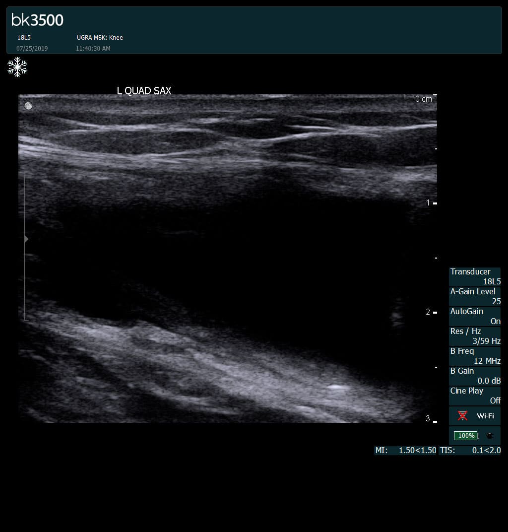
Complete tear of rectus femoris
with large hematoma (blood)
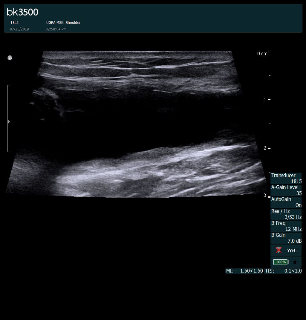
Separation of muscle ends due to tear elicited
on dynamic sonography examination


 Request Telehealth