October 27, 2025
Your ability to perform physical tasks and activities depends on a biomechanical lever system, where muscle pulls against bone at the joints to generate rotational torque that produces movement. The architecture of a particular joint dictates its range of motion, supported by tough inelastic ligaments that attach bone to bone, and by biotensegrity – a system of elastic tension generated by muscles and fascia that controls and guides forces that act at the joint.
Despite wide diversity in individual anatomy, most people share similar limitations in joint range of motion, enabling them to walk, run, jump, climb and lift with minimal risk of injury. But for people with hypermobile Ehlers-Danlos syndrome (hEDS), the tissues that provide joint support and dictate range of motion are more fragile and elastic, allowing for supranormal extensibility that makes joints prone to injury and limits participation in everyday physical activities.
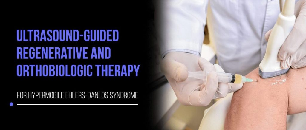
At NYDNRehab, we provide specialized hEDS physical therapy combined with evidence-based regenerative and orthobiologic approaches to strengthen fragile tissues, treat injuries, improve joint stability and manage pain symptoms. Learn about hEDS, the benefits of regenerative and orthobiologic therapies for treatment, and the importance of high-resolution ultrasound guidance for precise and gentle interventions.
Ehlers-Danlos syndrome is a group of hereditary connective tissue disorders that affect the skin, joints, nerves and blood vessels. Twelve of the 13 subtypes of EDS have clear genetic markers, but hypermobile EDS – the most common type – has no known genetic biomarkers and possesses its own systemic issues.
The most prominent features of hEDS are related to fragile connective tissues throughout the body, leading to a wide array of condition-specific symptoms. Diagnosis is based entirely on clinical criteria, as no genetic cause has yet been identified.
Characteristics of hEDS include some or all of the following:
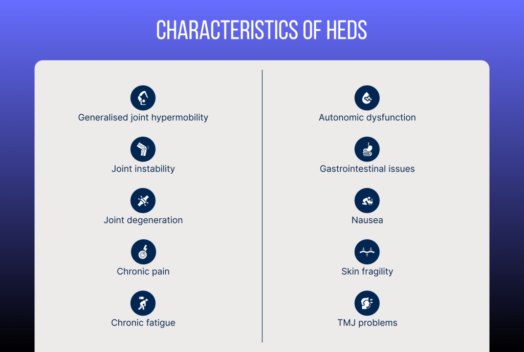
The prevalence of hEDS is not clearly known due to the complexities of diagnostic criteria. Certain people have hypermobile joints but are not affected by other hEDS symptoms. Some sources estimate hEDS to affect one in 3000-5000 people, translating to about .02-.03 percent of the population.
Patients with hEDS face serious quality of life issues affecting multiple facets of daily living. Pain and fatigue can affect consistency and quality of work, and many hEDS sufferers have difficulty maintaining a job. Participation in sports and exercise is limited due to low exercise tolerance and fear of triggering pain symptoms. Patients often suffer from depression and anxiety, and many limit their social interactions for fear of fainting or symptom flare-ups in public spaces.
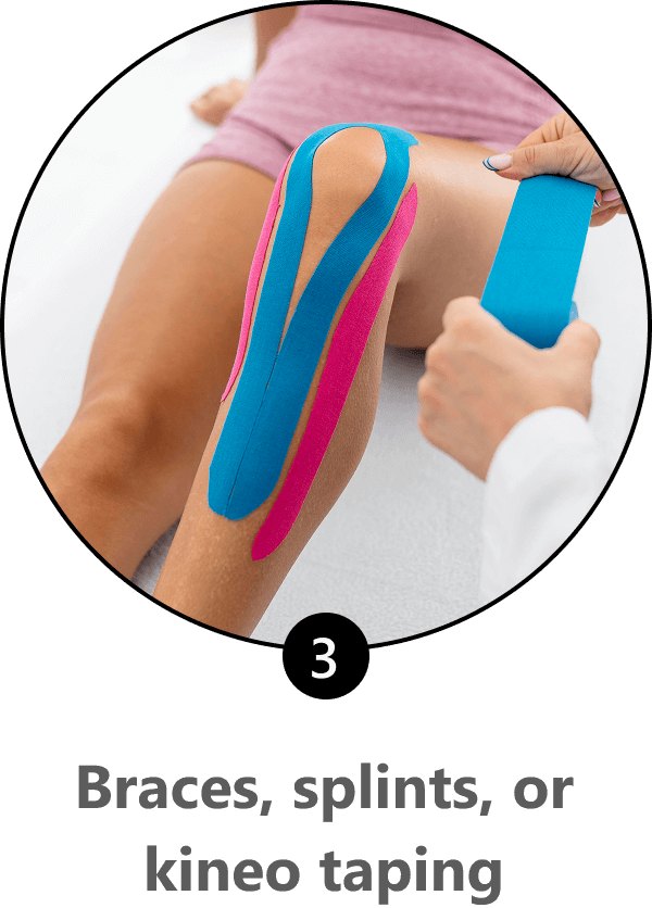
There is no cure for hEDS, and conventional treatments focus mainly on managing symptoms, improving function, and enhancing quality of life. Treatment approaches often include physical therapy, pain relief medications, taping and bracing for joint support, management of autonomic and GI symptoms, patient education, and mental health support.
Holistic therapy for hEDS incorporates some conventional interventions, but it typically steers clear of medications and leverages alternative therapies for pain management. Its whole-body approach emphasizes tissue healing, strength optimization, and restoration and maintenance of biotensegrity, with an emphasis on fascial health.
But symptoms management is not the ultimate goal. The end goal is to help hEDS patients achieve the best possible quality of life. Patients face significant quality of life challenges due to chronic pain, joint instability, fatigue, gastrointestinal issues, autonomic dysfunction, mental health struggles, and social/occupational limitations. These issues create a complex burden that requires personalized multidisciplinary management.
At NYDNRehab, our personalized approach to patient care means hEDS patients get the support and care they need to feel seen and validated. We leverage the most advanced energy technologies and orthobiologic procedures for hEDS therapy, supported by evidence from a growing body of research. Our successful adoption of cutting-edge approaches is forging new pathways for holistic hEDS treatment.
Hypermobile Ehlers-Danlos syndrome involves defective connective tissues, leading to joint hypermobility, chronic pain, instability, and tissue fragility. Regenerative therapies and orthobiologics target hEDS symptoms such as joint laxity, tendinopathies, and chronic pain, helping patients to avoid invasive surgery, which can be disastrous for hEDS sufferers.
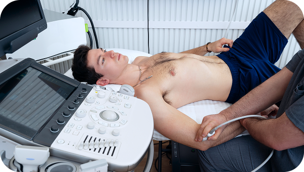
At NYDNRehab, we guide our regenerative and orthobiologic procedures with high-resolution ultrasonography, to ensure precision targeting of fragile tissues. With our advanced ultrasound equipment, we are able to adjust the wave type and depth to ensure precise and gentle interventions. Without ultrasound imaging, such procedures can be hit-or-miss, potentially doing more harm than good. Dr. Kalika’s expertise in ultrasonography sets NYDNRehab apart from other clinics offering similar services.
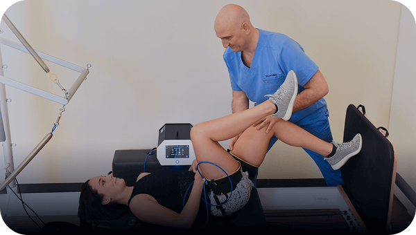
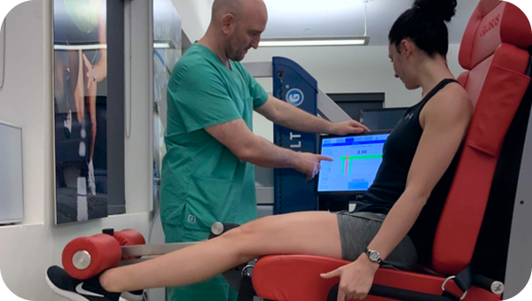
Regenerative technologies use acoustic, electric, magnetic and radiofrequency waves to stimulate tissue regeneration at the cellular level. At NYDNRehab, we carefully select a combination of technologies, based on the needs of the individual patient. Our personalized approach to hEDS ensures that you get appropriate and effective treatment that eases hEDS symptoms and promotes joint stability.
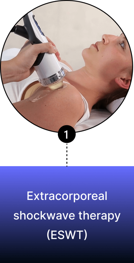
Extracorporeal shockwave therapy (ESWT)
ESWT uses high-energy acoustic waves that stimulate tissue healing. When applied at the appropriate depth, scope and intensity, ESWT can help to restore degenerated tendon tissues, improve ligament stability, enhance proprioception, and reduce chronic pain. It is especially effective for treating overuse injuries, microtears or adhesions in fascial and tendinous tissues. Patients often report relief after one to three sessions.
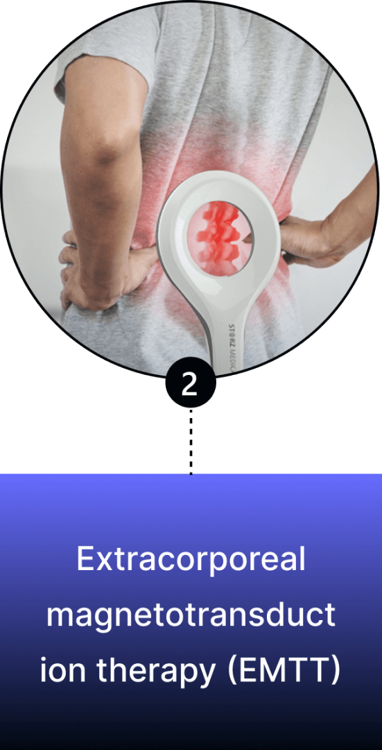
Extracorporeal magnetotransduction therapy (EMTT)
EMTT uses high-intensity magnetic fields that stimulate cellular repair, reduce inflammation, and promote regeneration in tendons, ligaments and joints. EMTT targets defective collagen that weakens tissues – a core issue in hEDS.
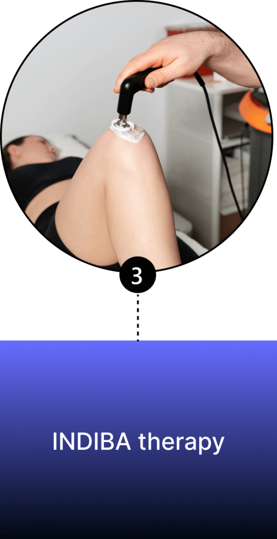
INDIBA therapy
INDIBA uses radiofrequency electromagnetic waves to generate controlled heat deep within tissues. INDIBA operates in two modes – capacitive, for superficial tissues like skin and muscles, and resistive, for deeper structures like tendons, ligaments, and bones. For hEDS, INDIBA enhances circulation, reduces muscle spasms, and supports tissue remodeling, with the goal of improving the stability and function of fragile joints.
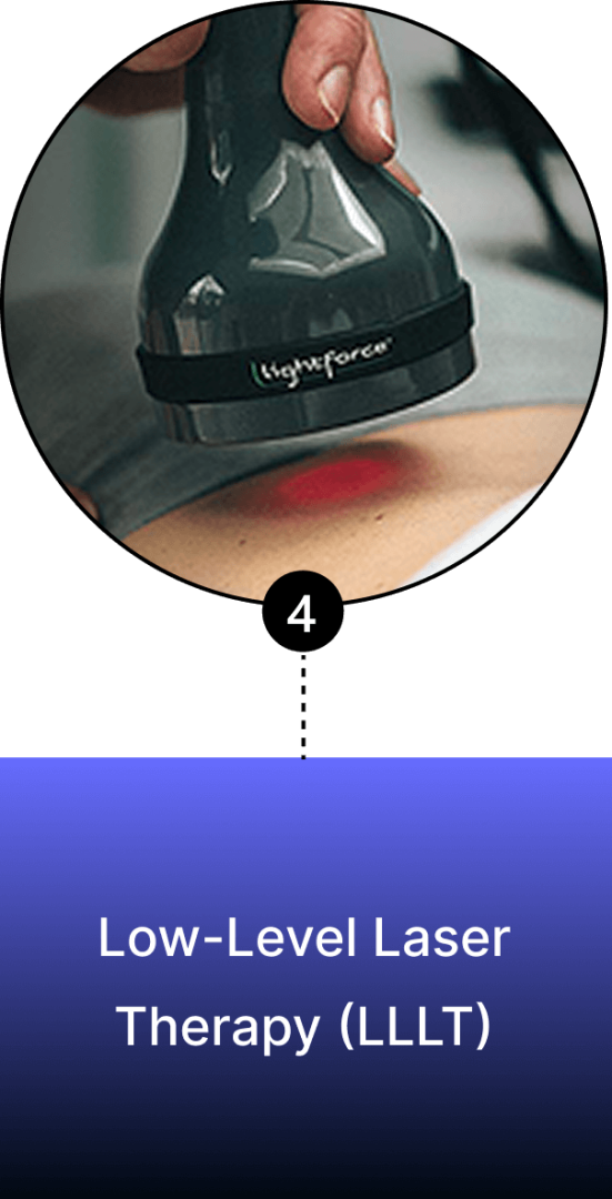
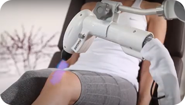
Low-Level Laser Therapy (LLLT)
Laser therapy uses low-intensity light to reduce chronic joint and muscle pain, enhance collagen synthesis in tendons and ligaments, improve joint range of motion, and reduce subluxations. It enhances blood circulation, easing symptoms of dysautonomia.
Orthobiologic procedures inject biological or neutral solutions into damaged tissues and joints to stimulate tissue repair at the cellular level, targeting symptoms like joint laxity, tendinopathies, and pain without invasive surgery. Dr. Kalika’s expertise in ultrasonography combined with Dr. Brosgol’s skills in orthobiologic procedures ensure that hEDS patients get targeted and gentle treatment that strengthens fragile tissues and reduces instability.
Prolotherapy
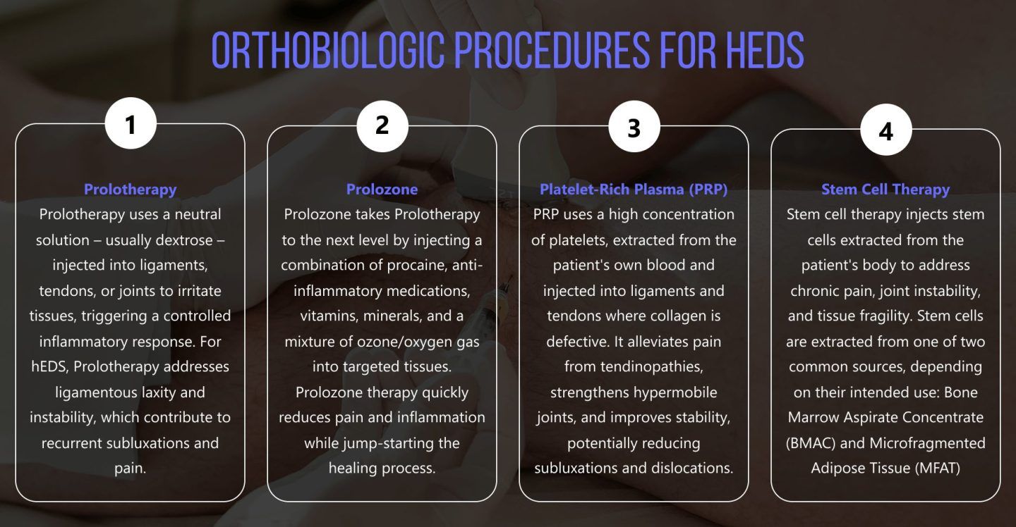
Prolotherapy uses a neutral solution – usually dextrose – injected into ligaments, tendons, or joints to irritate tissues, triggering a controlled inflammatory response. For hEDS, Prolotherapy addresses ligamentous laxity and instability, which contribute to recurrent subluxations and pain.
Platelet-Rich Plasma (PRP)
PRP uses a high concentration of platelets, extracted from the patient’s own blood and injected into ligaments and tendons where collagen is defective. It alleviates pain from tendinopathies, strengthens hypermobile joints, and improves stability, potentially reducing subluxations and dislocations.
Stem Cell Therapy
Stem cell therapy injects stem cells extracted from the patient’s body to address chronic pain, joint instability, and tissue fragility. Stem cells are extracted from one of two common sources, depending on their intended use:
At NYDNRehab, treatment for hEDS involves a combination of carefully selected therapies, based on the patient’s unique needs. When combined with specialized hEDS physical therapy, our holistic and personalized approach can dramatically improve joint stability and enhance patient quality of life.
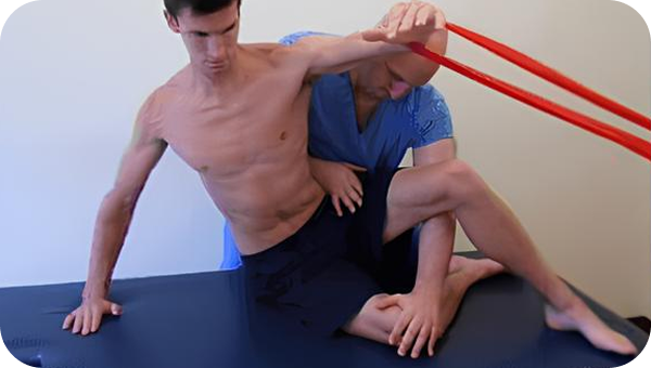
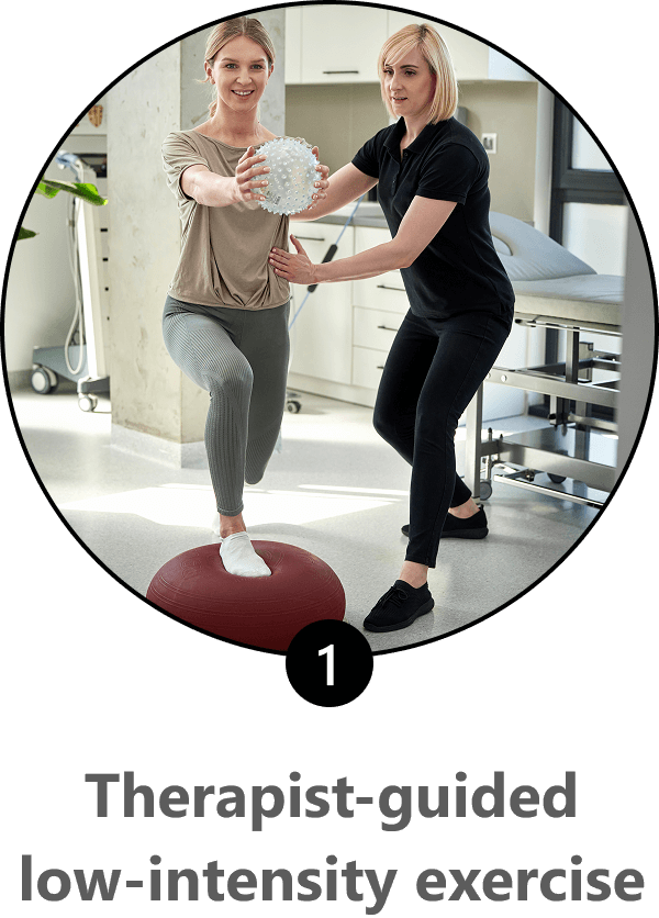
Conventional physical therapy is not enough to address the peculiar aspects of hEDS, and an inexperienced or underqualified therapist could potentially cause harm. At NYDNRehab, our holistic approach includes alternative interventions not commonly used in mainstream clinics. Our personalized one-on-one approach ensures that your fragile tissues and unstable joints are protected during your physical therapy sessions.
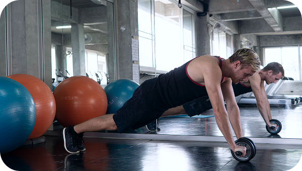
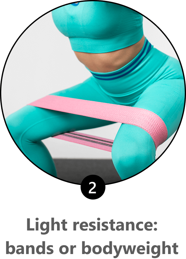
In addition to elastic resistance and body weight exercises, your hEDS physical therapy protocol may include:
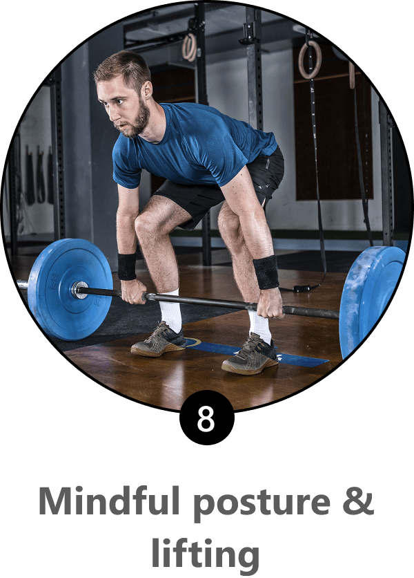
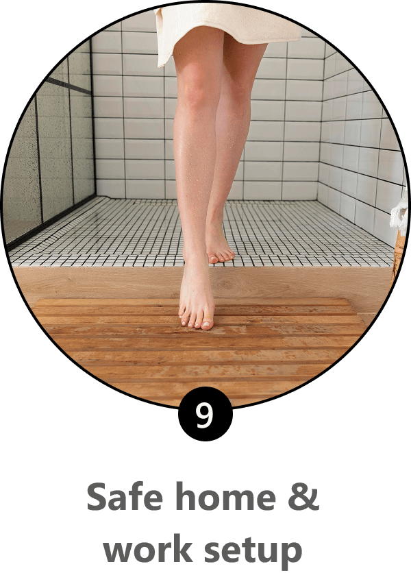
As with any chronic musculoskeletal condition, lifestyle factors go a long way toward managing and mitigating hEDS symptoms. The 2017 International Classification of the Ehlers–Danlos Syndromes provides the following lifestyle guidelines for hEDS injury prevention, centered on stabilizing joints, reducing strain, and preventing flare-ups. When integrated into daily life, these guidelines can significantly reduce injury risk and improve quality of life for hEDS sufferers.


Patients with hEDS face multiple daily hurdles that diminish their quality of life. When coupled with personalized hEDS physical therapy and supported by lifestyle modifications, regenerative therapies and orthobiologics can dramatically improve hEDS symptoms and reduce injury risk.
NYDNRehab is one of the few clinics in NYC that specializes in hEDS therapy. Our state-of-the-art clinic features a broad spectrum of technologies and treatment options, equipping us with the best tools for personalized hEDS care. To optimize your joint stability, enhance your tissue quality, and reduce hEDS symptoms, contact NYDNRehab today for holistic hEDS treatment without drugs or surgery.

Verified Expert Profiles
Dr. Lev Kalika is a world-recognized expert in musculoskeletal medicine. with 20+ years of clinical experience in diagnostic musculoskeletal ultrasonography, rehabilitative sports medicine and conservative orthopedics. In addition to operating his clinical practice in Manhattan, he regularly publishes peer-reviewed research on ultrasound-guided therapies and procedures. He serves as a peer reviewer for Springer Nature.
Dr. Kalika is an esteemed member of multiple professional organizations, including:
Below is a prime example of how ultrasound can take the guesswork out of diagnosis.

A bad physical therapy experience is one of the primary causes of unnecessary surgery
In this instance, an athlete was originally diagnosed with minor quadriceps muscle strain and was treated for four weeks, with unsatisfactory results. When he came to our clinic, the muscle was not healing, and the patients’ muscle tissue had already begun to atrophy.
Upon examination using MSUS, we discovered that he had a full muscle thickness tear that had been overlooked by his previous provider. To mitigate damage and promote healing, surgery should have been performed immediately after the injury occurred. Because of misdiagnosis and inappropriate treatment, the patient now has permanent damage that cannot be corrected.
The most important advantage of Ultrasound over MRI imaging is its ability to zero in on the symptomatic region and obtain imaging, with active participation and feedback from the patient. Using dynamic MSUS, we can see what happens when patients contract their muscles, something that cannot be done with MRI. From a diagnostic perspective, this interaction is invaluable.
Dynamic ultrasonography examination demonstrating
the full thickness tear and already occurring muscle atrophy
due to misdiagnosis and not referring the patient
to proper diagnostic workup
Demonstration of how very small muscle defect is made and revealed
to be a complete tear with muscle contraction
under diagnostic sonography (not possible with MRI)

Complete tear of rectus femoris
with large hematoma (blood)

Separation of muscle ends due to tear elicited
on dynamic sonography examination


