Our patient is a 31-year-old female experiencing knee pain during standing and walking. The patient presented with a Genu Valgum angle of 15-20 degrees.
In addition to a clinical exam and measuring the Genu Valgum angle, we used high-resolution diagnostic ultrasound to examine the muscles and connective tissues that act at the knee. We were able to recognize muscle activation patterns that were contributing to Genu Valgum and employed functional ultrasound to identify the affected muscle fibers
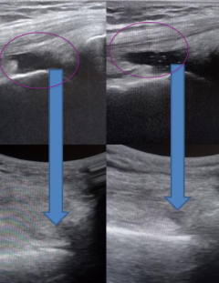
Our dynamic ultrasound exam identified abnormalities in the flexor digitorum longus and flexor digitorum brevis muscles of the calf that were affecting the patient’s knee angle. A functional ultrasound exam of the affected muscle fibers and the muscles on the plantar surface during toe movements revealed the presence of multiple myofascial trigger points.
We used precision dry needling under ultrasound guidance to eliminate myofascial trigger points that were creating muscle imbalances that misaligned the knee joints.
This case study underscores the role of ultrasound-guided dry needling in correcting adult Genu Valgum. Ultrasound guidance enhances precision of the dry needling procedure. Dynamic ultrasonography is also beneficial in identifying uncoordinated muscle activation patterns that affect knee alignment, and detecting affected muscle fibers.
Precise detection and treatment of miniscule trigger points, sometimes as small as 3 mm, requires a meticulous and skilled approach, and an in-depth knowledge of the interactions of muscles and their activation patterns. Advanced technologies coupled with experience and expertise allow for a personalized and targeted treatment approach.

Verified Expert Profiles
Dr. Lev Kalika is a world-recognized expert in musculoskeletal medicine. with 20+ years of clinical experience in diagnostic musculoskeletal ultrasonography, rehabilitative sports medicine and conservative orthopedics. In addition to operating his clinical practice in Manhattan, he regularly publishes peer-reviewed research on ultrasound-guided therapies and procedures. He serves as a peer reviewer for Springer Nature.
Dr. Kalika is an esteemed member of multiple professional organizations, including:
Below is a prime example of how ultrasound can take the guesswork out of diagnosis.

A bad physical therapy experience is one of the primary causes of unnecessary surgery
In this instance, an athlete was originally diagnosed with minor quadriceps muscle strain and was treated for four weeks, with unsatisfactory results. When he came to our clinic, the muscle was not healing, and the patients’ muscle tissue had already begun to atrophy.
Upon examination using MSUS, we discovered that he had a full muscle thickness tear that had been overlooked by his previous provider. To mitigate damage and promote healing, surgery should have been performed immediately after the injury occurred. Because of misdiagnosis and inappropriate treatment, the patient now has permanent damage that cannot be corrected.
The most important advantage of Ultrasound over MRI imaging is its ability to zero in on the symptomatic region and obtain imaging, with active participation and feedback from the patient. Using dynamic MSUS, we can see what happens when patients contract their muscles, something that cannot be done with MRI. From a diagnostic perspective, this interaction is invaluable.
Dynamic ultrasonography examination demonstrating
the full thickness tear and already occurring muscle atrophy
due to misdiagnosis and not referring the patient
to proper diagnostic workup
Demonstration of how very small muscle defect is made and revealed
to be a complete tear with muscle contraction
under diagnostic sonography (not possible with MRI)

Complete tear of rectus femoris
with large hematoma (blood)

Separation of muscle ends due to tear elicited
on dynamic sonography examination


