A 33-year-old female came to us complaining of persistent left groin pain and painful intercourse. She had been seeing a physical therapist for hip pain that extended to her groin and pelvis.
In addition to physical therapy, the patient was treated with manual internal pelvic floor massage. When her condition did not improve, she came to our clinic for further diagnosis and treatment.
The patient’s health history revealed that she had undergone an abdominal laparoscopy five years prior. The clinical exam revealed no specific musculoskeletal issues. We performed rehabilitative functional ultrasound (RUSI) to observe the movement and contraction of her pelvic floor muscles, but did not visualize any abnormalities.
We referred the patient to a gynecologist who did not find anything, and who ruled out the possibility of an inguinal hernia.
Upon the patient’s return to us, we palpated her abdomen, pelvis and groin to see if we could reproduce her pain, and also to see if her prior laparoscopy had caused abnormal intra abdominal thickening or abdominal fascial distortion. We found some minor densifications along the ante-medial line that did not reproduce her pain, but there was some localized tenderness.
We then used a curvilinear probe to examine peristaltic movement via ultrasound, to rule out visceral adhesions. We visualized an area of increased echogenicity near the sigmoid colon, so we sent the patient to her primary care physician to request a referral for abdominal ultrasound or a CT scan.
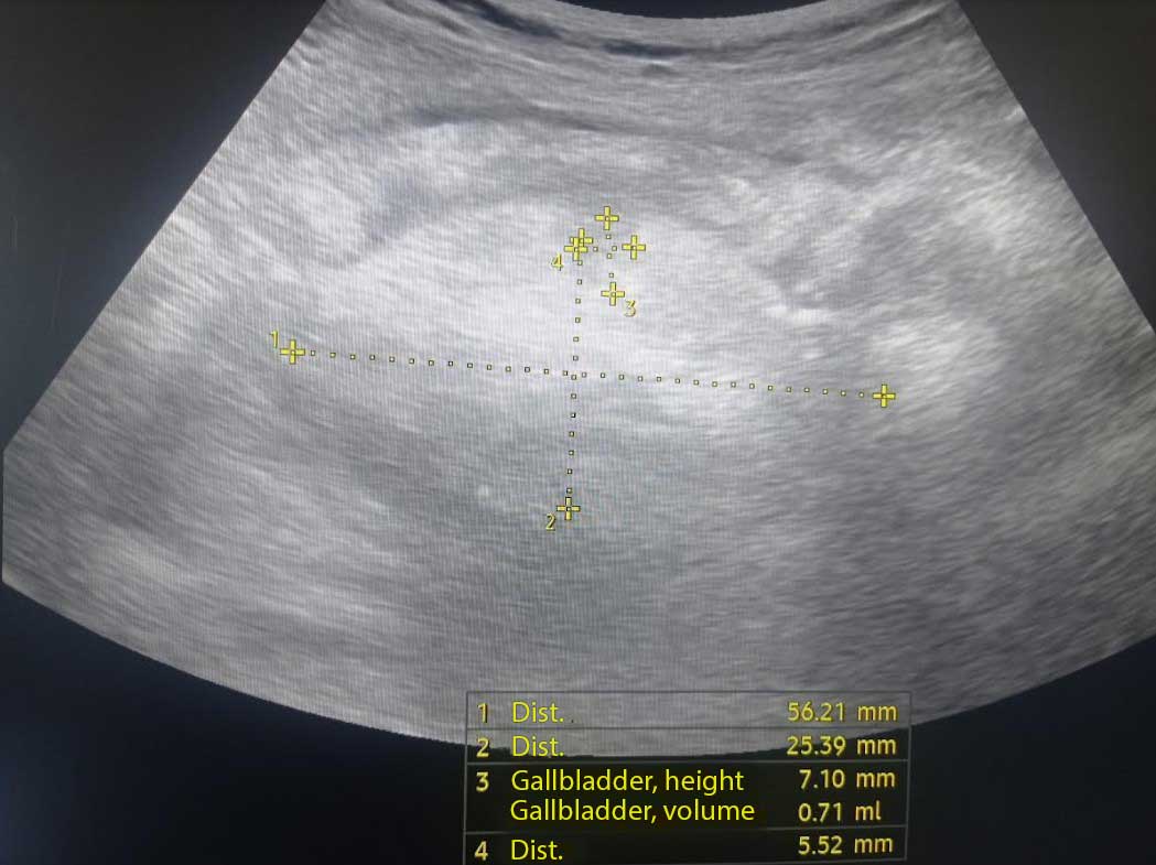
An abdominal ultrasound revealed significant findings:
Based on our findings, the patient was diagnosed with sigmoiditis, a non-muscular chronic inflammation of the colon that was causing her persistent left groin pain.
Given the non-musculoskeletal nature of the patient’s condition, we recommended that physical therapy should primarily focus on any potential complications related to sigmoiditis, as well as managing any associated symptoms. Collaboration with the patient’s primary healthcare provider would be crucial to ensure a comprehensive and coordinated treatment plan.
Many therapists are quick to assume that pelvic pain is caused by overly tight pelvic floor muscles, and that stretching the muscles via manual manipulation is a catch-all solution. However, it is important to consider that the pelvis houses multiple non-muscular structures that may also be a source of pelvic pain and dysfunction. Moreover, manual stretching of the pelvic floor muscles has scant scientific evidence to support it, despite its widespread use.

Verified Expert Profiles
Dr. Lev Kalika is a world-recognized expert in musculoskeletal medicine. with 20+ years of clinical experience in diagnostic musculoskeletal ultrasonography, rehabilitative sports medicine and conservative orthopedics. In addition to operating his clinical practice in Manhattan, he regularly publishes peer-reviewed research on ultrasound-guided therapies and procedures. He serves as a peer reviewer for Springer Nature.
Dr. Kalika is an esteemed member of multiple professional organizations, including:
Below is a prime example of how ultrasound can take the guesswork out of diagnosis.

A bad physical therapy experience is one of the primary causes of unnecessary surgery
In this instance, an athlete was originally diagnosed with minor quadriceps muscle strain and was treated for four weeks, with unsatisfactory results. When he came to our clinic, the muscle was not healing, and the patients’ muscle tissue had already begun to atrophy.
Upon examination using MSUS, we discovered that he had a full muscle thickness tear that had been overlooked by his previous provider. To mitigate damage and promote healing, surgery should have been performed immediately after the injury occurred. Because of misdiagnosis and inappropriate treatment, the patient now has permanent damage that cannot be corrected.
The most important advantage of Ultrasound over MRI imaging is its ability to zero in on the symptomatic region and obtain imaging, with active participation and feedback from the patient. Using dynamic MSUS, we can see what happens when patients contract their muscles, something that cannot be done with MRI. From a diagnostic perspective, this interaction is invaluable.
Dynamic ultrasonography examination demonstrating
the full thickness tear and already occurring muscle atrophy
due to misdiagnosis and not referring the patient
to proper diagnostic workup
Demonstration of how very small muscle defect is made and revealed
to be a complete tear with muscle contraction
under diagnostic sonography (not possible with MRI)

Complete tear of rectus femoris
with large hematoma (blood)

Separation of muscle ends due to tear elicited
on dynamic sonography examination


