Our patient is a 33 year-old female runner complaining of hamstring and calf tightness, and posterior knee pain in both legs. The pain began in 2015, but she continued to run.
The patient had seen numerous running physical therapists and had undergone multiple gait analyses without any results. Five years prior she had sustained tears in the medial collateral ligament (MCL) and posterior cruciate ligament (PCL) in the right knee when windsurfing.
We conducted a 3D gait analysis using advanced technologies and proprietary software to get an objective picture of the patient’s gait mechanics. Gait analysis results revealed multiple issues:
We conducted a battery of tests and assessments to evaluate posture and biomechanics. We determined that the patient:
We used high-resolution diagnostic ultrasound to visualize the patient’s right knee:
When conducted by an experienced clinician who can clinically interpret results, 3D gait analysis can be an extremely useful tool. However, even the most advanced gait analysis system cannot always explain the reasons for mechanical deficits. The data produced tells us what is wrong, but we still have to figure out the underlying cause, and differentiate compensation patterns from pathology. Diagnosis requires clinical experience, and expertise with other diagnostic tools such as high resolution diagnostic ultrasonography.

Verified Expert Profiles
Dr. Lev Kalika is a world-recognized expert in musculoskeletal medicine. with 20+ years of clinical experience in diagnostic musculoskeletal ultrasonography, rehabilitative sports medicine and conservative orthopedics. In addition to operating his clinical practice in Manhattan, he regularly publishes peer-reviewed research on ultrasound-guided therapies and procedures. He serves as a peer reviewer for Springer Nature.
Dr. Kalika is an esteemed member of multiple professional organizations, including:
Below is a prime example of how ultrasound can take the guesswork out of diagnosis.

A bad physical therapy experience is one of the primary causes of unnecessary surgery
In this instance, an athlete was originally diagnosed with minor quadriceps muscle strain and was treated for four weeks, with unsatisfactory results. When he came to our clinic, the muscle was not healing, and the patients’ muscle tissue had already begun to atrophy.
Upon examination using MSUS, we discovered that he had a full muscle thickness tear that had been overlooked by his previous provider. To mitigate damage and promote healing, surgery should have been performed immediately after the injury occurred. Because of misdiagnosis and inappropriate treatment, the patient now has permanent damage that cannot be corrected.
The most important advantage of Ultrasound over MRI imaging is its ability to zero in on the symptomatic region and obtain imaging, with active participation and feedback from the patient. Using dynamic MSUS, we can see what happens when patients contract their muscles, something that cannot be done with MRI. From a diagnostic perspective, this interaction is invaluable.
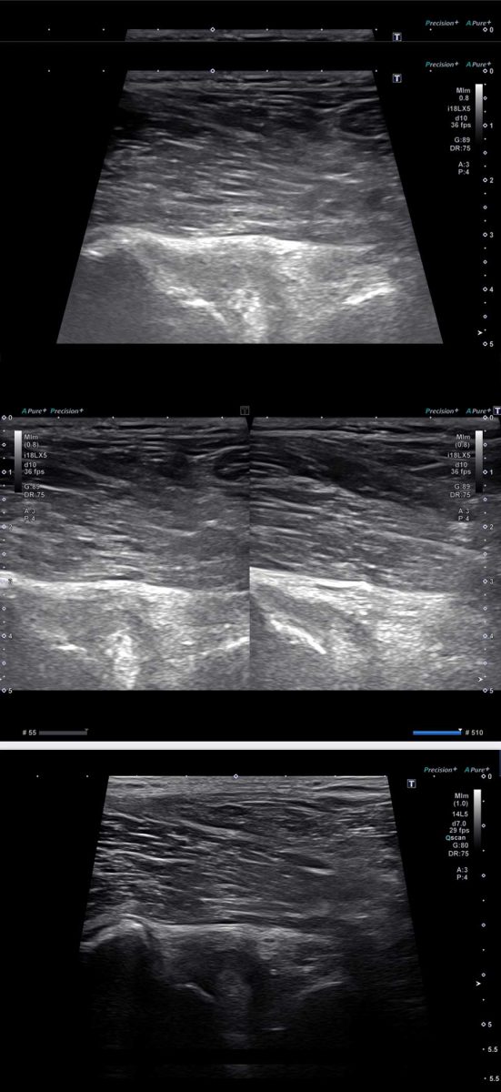
Dynamic ultrasonography examination demonstrating
the full thickness tear and already occurring muscle atrophy
due to misdiagnosis and not referring the patient
to proper diagnostic workup
Demonstration of how very small muscle defect is made and revealed
to be a complete tear with muscle contraction
under diagnostic sonography (not possible with MRI)

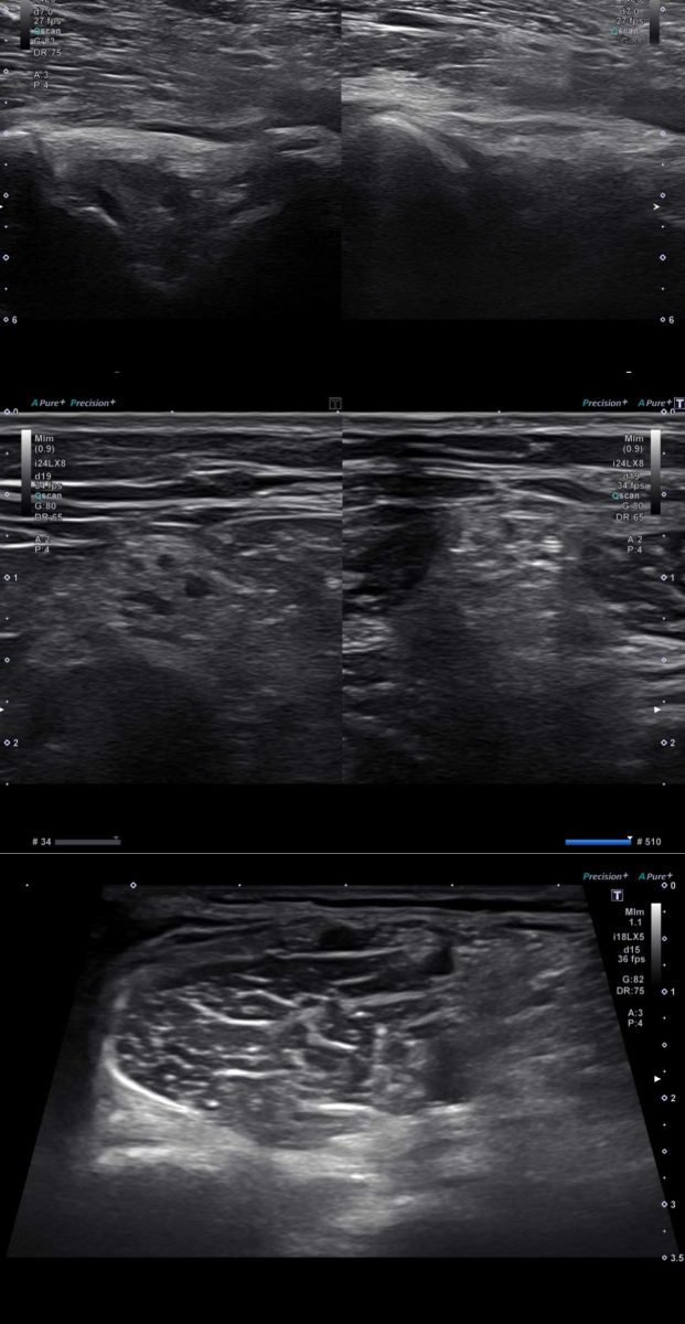
Complete tear of rectus femoris
with large hematoma (blood)

Separation of muscle ends due to tear elicited
on dynamic sonography examination


