Our patient was a 40-year-old female with bilateral knee hydrarthrosis (fluid accumulation) and shoulder impingement, who was suspected to have arthritis. Ultrasound imaging revealed no significant structural abnormalities in the knees. However, fluid accumulation was evident, particularly in the left knee.
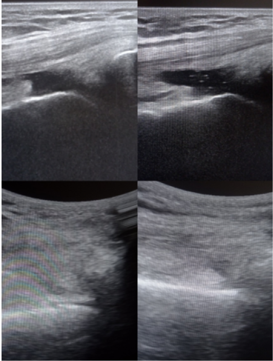
Accumulation of fluid detected in both knees.
In addition to excess knee fluid, ultrasound revealed trigger points in the right sacroiliac joint (SIJ) and multifidus muscles at L4-5, L2-4, and L, and in the left soleus muscle and medial quadriceps. The patient also had a history of thyroiditis bronchiectatic disease (inflamed thyroid and thickened bronchial walls), which was being treated with a steroid inhalant.
Our treatment plan included the use of ultrasound-guided dry needling to eliminate trigger points, which were contributing to mechanical overload on the joints and surrounding tissues, and causing postural misalignment. Postural correction techniques were used to restore functional alignment.
Our treatment had the effect of immediately reducing pain and eliminating fluid in both knees, while enhancing posture and completely resolving the patient’s shoulder impingement. The ability to precisely detect and eliminate trigger points using ultrasound guidance was key to resolving the patient’s mechanical abnormalities that were the source of pain and inflammation.
Our treatment approach supports the use of ultrasound-guided dry needing to manage joint fluid accumulation, eliminate pain, resolve mechanical overload, improve posture and restore functional mobility in patients with myofascial trigger points.

Verified Expert Profiles
Dr. Lev Kalika is a world-recognized expert in musculoskeletal medicine. with 20+ years of clinical experience in diagnostic musculoskeletal ultrasonography, rehabilitative sports medicine and conservative orthopedics. In addition to operating his clinical practice in Manhattan, he regularly publishes peer-reviewed research on ultrasound-guided therapies and procedures. He serves as a peer reviewer for Springer Nature.
Dr. Kalika is an esteemed member of multiple professional organizations, including:
Below is a prime example of how ultrasound can take the guesswork out of diagnosis.

A bad physical therapy experience is one of the primary causes of unnecessary surgery
In this instance, an athlete was originally diagnosed with minor quadriceps muscle strain and was treated for four weeks, with unsatisfactory results. When he came to our clinic, the muscle was not healing, and the patients’ muscle tissue had already begun to atrophy.
Upon examination using MSUS, we discovered that he had a full muscle thickness tear that had been overlooked by his previous provider. To mitigate damage and promote healing, surgery should have been performed immediately after the injury occurred. Because of misdiagnosis and inappropriate treatment, the patient now has permanent damage that cannot be corrected.
The most important advantage of Ultrasound over MRI imaging is its ability to zero in on the symptomatic region and obtain imaging, with active participation and feedback from the patient. Using dynamic MSUS, we can see what happens when patients contract their muscles, something that cannot be done with MRI. From a diagnostic perspective, this interaction is invaluable.
Dynamic ultrasonography examination demonstrating
the full thickness tear and already occurring muscle atrophy
due to misdiagnosis and not referring the patient
to proper diagnostic workup
Demonstration of how very small muscle defect is made and revealed
to be a complete tear with muscle contraction
under diagnostic sonography (not possible with MRI)

Complete tear of rectus femoris
with large hematoma (blood)

Separation of muscle ends due to tear elicited
on dynamic sonography examination


