Our patient is a 43 year-old female suffering from hand numbness and impingement of the right shoulder consequent to chemotherapy treatment for thymoma. The patient had previously sought medical attention at another clinic where her condition had been misdiagnosed, resulting in unsuccessful, costly and time-consuming treatment.
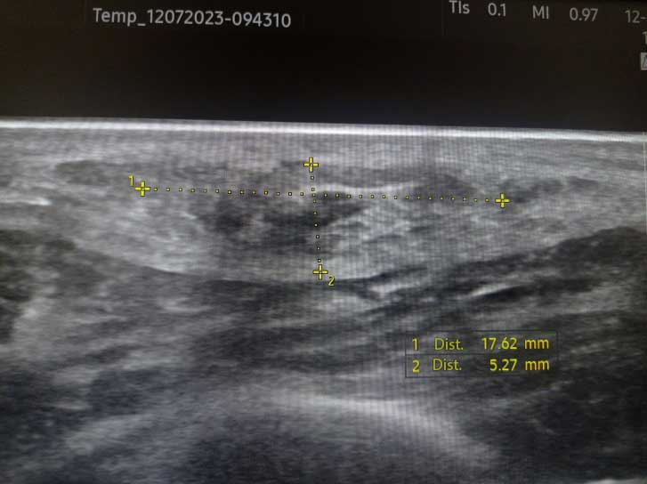
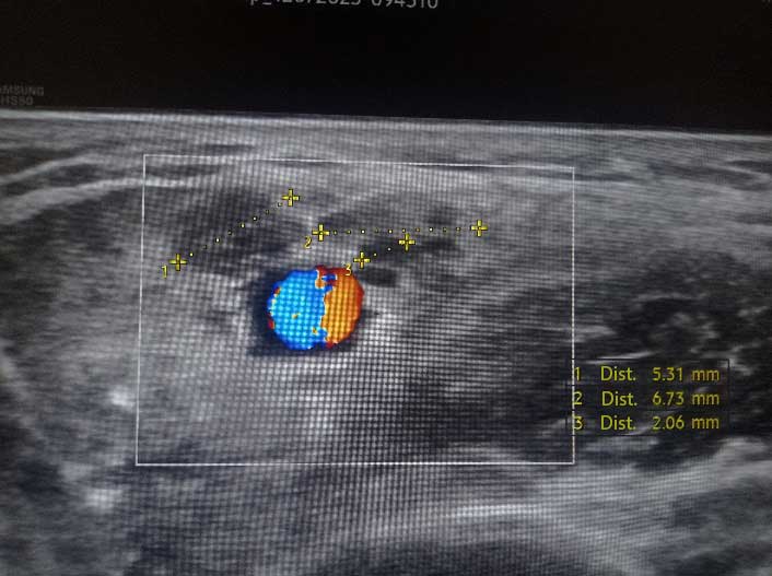
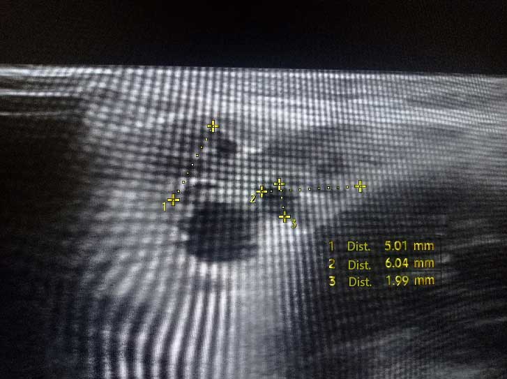
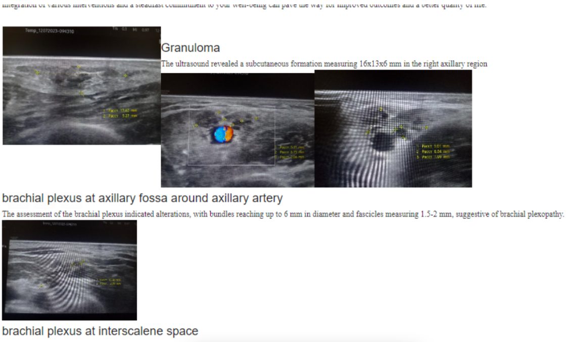
Our multidisciplinary team leveraged advanced ultrasound diagnostics to identify underlying factors contributing to the patient’s condition. We discovered the presence of axillary lymphadenopathy – an abnormality of the lymph node in the right armpit, marked by a granuloma of white blood cells. We also uncovered indicators of brachial plexopathy – a form of peripheral neuropathy affecting the nerves of the brachial plexus.
Given the patient’s previous misdiagnosis, our team wanted to ensure that our treatment plan provided both immediate relief and a strategy for long-term recovery. The following interdisciplinary measures were taken:
As a result of our multidisciplinary treatment approach, the patient’s symptoms significantly improved over time, and arm and shoulder functionality was significantly enhanced. Of particular benefit was ultrasound-guided dry needling (USGDN), which effectively addressed the brachial plexopathy and contributed to the patient’s overall recovery.

Verified Expert Profiles
Dr. Lev Kalika is a world-recognized expert in musculoskeletal medicine. with 20+ years of clinical experience in diagnostic musculoskeletal ultrasonography, rehabilitative sports medicine and conservative orthopedics. In addition to operating his clinical practice in Manhattan, he regularly publishes peer-reviewed research on ultrasound-guided therapies and procedures. He serves as a peer reviewer for Springer Nature.
Dr. Kalika is an esteemed member of multiple professional organizations, including:
Below is a prime example of how ultrasound can take the guesswork out of diagnosis.

A bad physical therapy experience is one of the primary causes of unnecessary surgery
In this instance, an athlete was originally diagnosed with minor quadriceps muscle strain and was treated for four weeks, with unsatisfactory results. When he came to our clinic, the muscle was not healing, and the patients’ muscle tissue had already begun to atrophy.
Upon examination using MSUS, we discovered that he had a full muscle thickness tear that had been overlooked by his previous provider. To mitigate damage and promote healing, surgery should have been performed immediately after the injury occurred. Because of misdiagnosis and inappropriate treatment, the patient now has permanent damage that cannot be corrected.
The most important advantage of Ultrasound over MRI imaging is its ability to zero in on the symptomatic region and obtain imaging, with active participation and feedback from the patient. Using dynamic MSUS, we can see what happens when patients contract their muscles, something that cannot be done with MRI. From a diagnostic perspective, this interaction is invaluable.
Dynamic ultrasonography examination demonstrating
the full thickness tear and already occurring muscle atrophy
due to misdiagnosis and not referring the patient
to proper diagnostic workup
Demonstration of how very small muscle defect is made and revealed
to be a complete tear with muscle contraction
under diagnostic sonography (not possible with MRI)

Complete tear of rectus femoris
with large hematoma (blood)

Separation of muscle ends due to tear elicited
on dynamic sonography examination


