Chronic ankle pain from an untreated injury can cause serious health problems, since healthy feet and ankles are foundational to all physical activity. Ankle pain can cause mechanical issues throughout your body, and can also impact your cardiovascular and metabolic health due to reduced activity.
or
Forty percent of people who sprain an ankle will develop chronic ankle instability (CAI), which will later develop into ankle osteoarthritis, for which there is currently no successful solution. Ankle replacement surgery is much more complicated and much less effective than knee or hip replacement. Regenerative procedures are also much less promising for repair of ankle cartilage damage, compared to knee or hip cartilage.
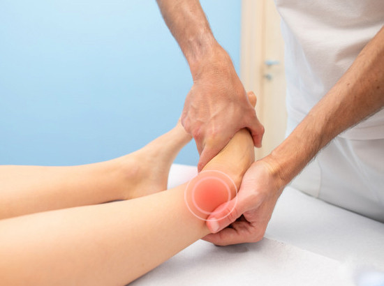
Before your undertreated ankle sprain begins to cause chronic ankle pain or turns into osteoarthritis, it will potentially destroy your hips, knees or low back without your awareness. Unfortunately, traditional medicine does not recognize the detrimental impact of ankle sprains on other body structures.
The problem is that doctors do not recognize problems associated with ankle sprains until it is too late, and many physios don’t know how to treat them, or have the right tools to do so.
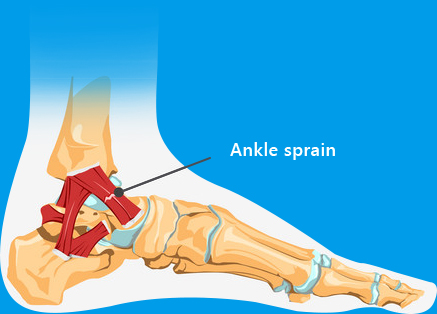


Many neurological and orthopedic changes are taking place in an unstable ankle. In fact, ankle sprains could aptly be renamed “brainkle” sprains, because they involve the neuromuscular system. These changes are intertwined and can only be reversed if all are addressed. They include:
I am proud to say that our clinic at NYDNRehab is the top rehab clinic for chronic ankle sprain treatment in the country. Many professional athletes from the US and abroad come to us to rehab their ankles. We have developed a ground-breaking methodology and acquired research-grade technologies to address the impairments that cause ankle instability.
In addition to conventional physical therapy, we use the highest available resolution ultrasonography for dynamic ligament imaging and needle guidance. Other treatments include:
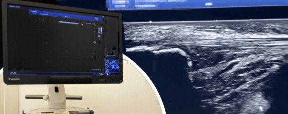
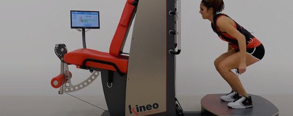

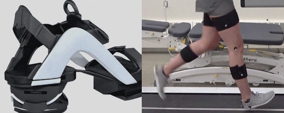

Dr.Kalika revolutionized foot and ankle care by using high resolution diagnostic ultrasonography for structural diagnosis, combined with with gait and motion analysis technology. Dr.Kalika’s motion and gait analysis lab is the only private lab in the US that features research-grade technology found only at top research universities, made available to patients in his private clinic.
 Dr. Yuri Brosgol
MD
Dr. Yuri Brosgol
MD
 Dr. Michael Goynatsky
DPT
Dr. Michael Goynatsky
DPT
 Dr. Daniela Escudero
DPT
Dr. Daniela Escudero
DPT
 Dr. Michelle Agyakwah
DC
Dr. Michelle Agyakwah
DC
 Dr. Tatyana Kapustina
L. Ac.
Dr. Tatyana Kapustina
L. Ac.

Below is a prime example of how ultrasound can take the guesswork out of diagnosis.

A bad physical therapy experience is one of the primary causes of unnecessary surgery
In this instance, an athlete was originally diagnosed with minor quadriceps muscle strain and was treated for four weeks, with unsatisfactory results. When he came to our clinic, the muscle was not healing, and the patients’ muscle tissue had already begun to atrophy.
Upon examination using MSUS, we discovered that he had a full muscle thickness tear that had been overlooked by his previous provider. To mitigate damage and promote healing, surgery should have been performed immediately after the injury occurred. Because of misdiagnosis and inappropriate treatment, the patient now has permanent damage that cannot be corrected.
The most important advantage of Ultrasound over MRI imaging is its ability to zero in on the symptomatic region and obtain imaging, with active participation and feedback from the patient. Using dynamic MSUS, we can see what happens when patients contract their muscles, something that cannot be done with MRI. From a diagnostic perspective, this interaction is invaluable.
Dynamic ultrasonography examination demonstrating
the full thickness tear and already occurring muscle atrophy
due to misdiagnosis and not referring the patient
to proper diagnostic workup
Demonstration of how very small muscle defect is made and revealed
to be a complete tear with muscle contraction
under diagnostic sonography (not possible with MRI)

Complete tear of rectus femoris
with large hematoma (blood)

Separation of muscle ends due to tear elicited
on dynamic sonography examination


