Physically active people are well aware of the role of muscle in human movement, and the majority of physical training is geared toward increasing muscle size, strength and power. What is poorly understood is that muscle does not act alone in force production – it works in concert with fascia and the nervous system to guide, control and distribute forces. Yet the synergistic relationship between muscle and fascia is often overlooked in conventional physical therapy. Treating injured muscles and joints while neglecting fascia can leave the patient with unresolved issues that continue to cause pain and dysfunction.
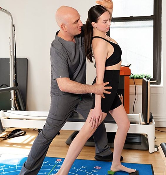
The Stecco technique of fascial manipulation is a highly systematic approach that requires specific training. Unlike other manual myofascial therapies, when applied correctly, Stecco Fascial Manipulation not only restores tissue gliding – it reactivates sensory receptors within the fascia, recalibrating the neuromuscular system and restoring efficient motor control. The Stecco technique also influences the neurohumeral, paracrine, hormonal, and visceral-somatic systems, supporting whole body regulation and recovery. When combined with regenerative and orthobiologic solutions, Stecco therapy can make the difference between partial and full recovery.
Regenerative therapies, orthobiologic procedures, and advanced manual techniques are opening new frontiers in rehabilitative medicine, with promising advancements on the horizon.
Clinical director & DC RMSK
Verified Expert Profiles
Dr. Lev Kalika, DC clinical director of NYDNRehab, is an internationally recognized expert in diagnostic and musculoskeletal ultrasound imaging, with multiple research papers to his credit. Dr. Kalika has studied with some of the world’s most prestigious experts in diagnostic, fascia, and nerve ultrasonography, and has presented his research at multiple international professional conferences.
Dr. Kalika is an active member of the American Institute of Ultrasound in Medicine (AIUM), and has developed his own unique approach to Dynamic Functional and Fascial Ultrasonography.

Orthobiologic, Sports Medicine and Regenerative Medicine Specialist
Muscle fibers contract (shorten) in response to neural signaling from the brain to the motor unit – a segment of muscle tissue defined by one motor neuron and the muscle fibers it innervates. When the brain sends a message to the motor neuron, all of its associated fibers contract simultaneously – a phenomenon called the “all or nothing” principle of muscle contraction.
The number of muscle fibers per motor unit varies widely throughout the body, depending on their purpose and location. Generally speaking, small motor units responsible for precise, controlled movements such as those of the eye and hand muscles, may have only 10–100 muscle fibers per motor unit. By contrast, larger muscles like the quadriceps or glutes may have thousands of muscle fibers per motor unit, capable of generating powerful forces, but with less precision.
Muscles and nerves do not act alone. Muscle actions are controlled and guided with the help of fascia, a complex web of thin, tough connective tissue that surrounds and connects muscles, engulfs the visceral organs, and holds the body’s structures in place during dynamic movement. Fascia is strong and elastic, able to stretch and contract as the body moves, providing elastic tension that complements muscle action.
Fascia itself is densely embedded with sensory nerve endings, including proprioceptors, nociceptors, and mechanoreceptors – neural bodies that send feedback to the brain about the body’s position in space, as well as sensations of pain and pressure. Fascia is estimated to have over 250 million nerve endings, making it a major pain generator when injured. Fascia, muscles and nerves act synergistically to inform the brain of the body’s state at any given time.
Fascia is made up of collagen fibers, lubricated by hyaluronic acid – a slippery gel-like substance able to attract 100X its mass in water. In addition to its elastic and neural properties, facia provides a slippery surface that allows nerves and blood vessels to glide freely among larger structures while allowing larger structures to move in harmony, without friction.
When fascia is injured, it can become dense and sticky, losing its elastic and slippery properties and adhering to other structures. Nerves and blood vessels can become entrapped in densified fascia, causing pain and restricting movement. Muscle action can be impaired, disrupting motor unit recruitment patterns and reducing movement efficiency.
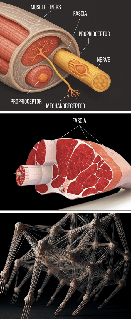
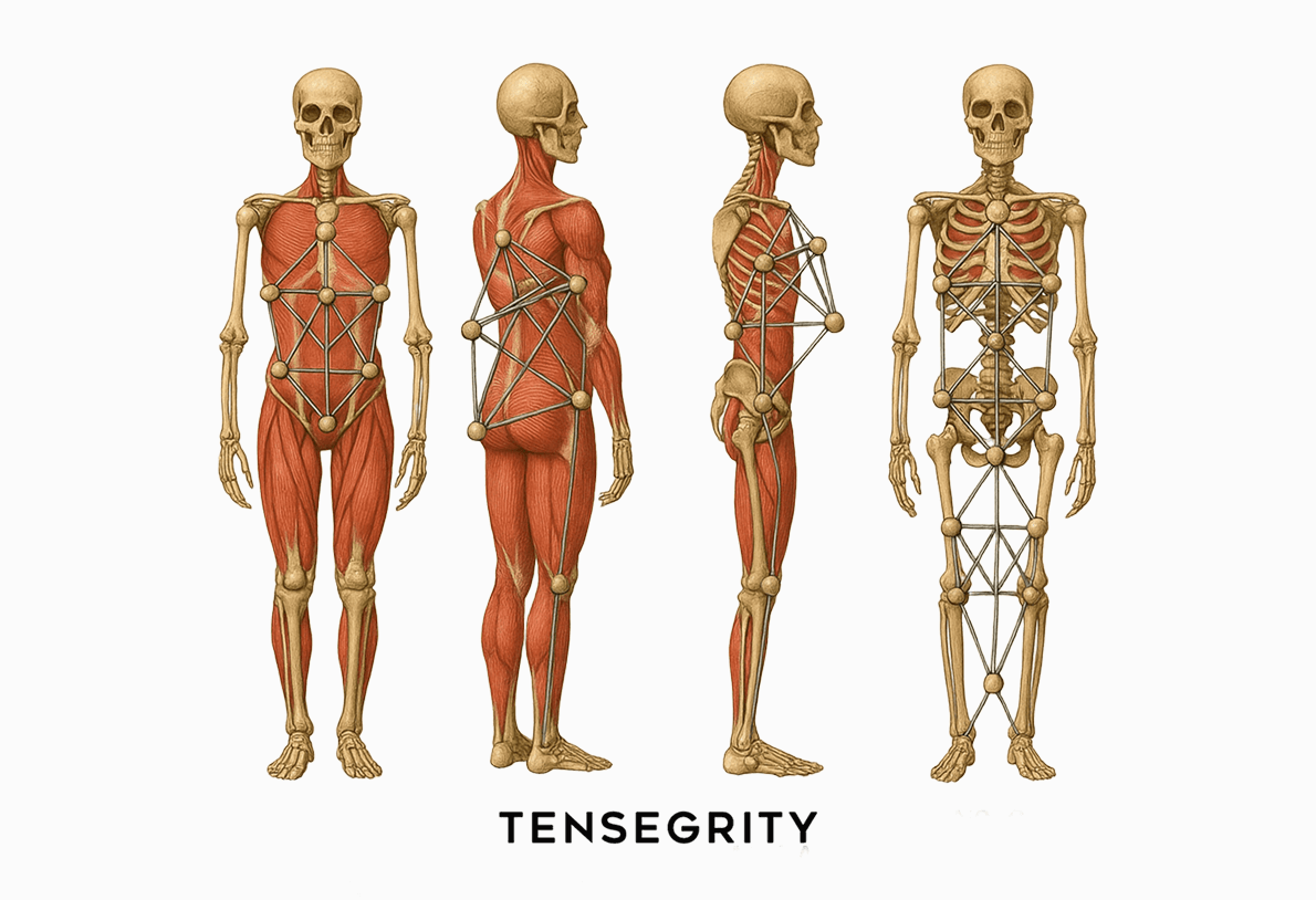
The myofascial system includes muscle and fascia, and their associated neural bodies. During physical activity, muscles and fascia work together to provide tensile integrity – aka biotensegrity – a state of elastic tension that guides and controls movement, holds organs and other structures in place, and mediates outside forces. When muscles, fascia or both are damaged, biotensegrity is compromised and mobility is impaired.
Restoring biotensegrity is a key factor in injury rehabilitation. It is not enough for injured muscles tissues to heal and pain to subside. Restoring the elastic and slippery properties of fascia is a critical step in injury rehab that should be addressed early on, before physical therapy begins. Unless the damaged fascial layers are treated and restored, mobility will continue to be impaired, increasing the risk of future injuries.
At NYDNRehab, we pre-treat injured tissues before beginning physical therapy. Our end goal is to fully restore functional mobility, and that cannot be achieved without addressing biotensegrity. Starting physical therapy prematurely can result in further tissue damage, and it can reinforce inefficient motor patterns that undermine physical performance.
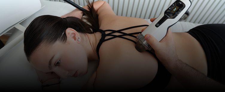
To treat densified fascia and fascial adhesions, multiple approaches have emerged in physical therapy, sports and fitness, such as foam rolling, myofascial massage, cupping, and other therapies aimed at releasing restrictions and alleviating pain. But while those techniques may provide some relief, they often fall short of fully restoring fascia’s functional properties.
Unlike other manual therapies, the Stecco technique of fascial manipulation is a highly systematic approach that requires specific training. When applied correctly, Stecco Fascial Manipulation not only restores tissue gliding – it reactivates sensory receptors within the fascia, helping the body to reorganize motor control, recalibrating the neuromuscular system and restoring efficient motor control.
Such reorganization occurs not only at the local (receptor and muscle) level, but throughout the entire nervous system, promoting more coordinated and efficient movement patterns. In addition, Stecco Fascial Manipulation influences the neurohumeral, paracrine, hormonal, and visceral-somatic systems, supporting global body regulation and recovery.
Progression of Stecco Fascial Manipulation involves:
These adaptations occur over the course of a week or two as tissue integrity and neural pathways are restored. This advanced methodology works to alleviate pain, eliminate restricted movement, enhance physical performance, and optimize whole-body mobility and stability. Dr. Kalika has received advanced training directly from Dr. Carla Stecco, creator of the Stecco Method and the world’s leading specialist in fascial science.
Mainstream physical rehab typically relies on standardized treatment protocols and estimated recovery timelines that treat all injuries alike. Treatment is often focused on the locus of pain, with little regard for adjacent and distal structures that may also be affected. Failure to consider the full spectrum of potential damage can leave you with lingering issues that manifest later on, impairing your performance and increasing your risk of injury.
Our holistic and integrative approach to injury rehab sets NYDNRehab apart from mainstream clinics. We treat the whole patient, not just your symptoms. We never take a one-size-fits-all approach to physical rehab. Our one-on-one sessions ensure that you get the full attention of your therapist, so we can fine-tune your rehab program as you heal.
We closely monitor your progress to ensure that your personalized protocol is delivering its intended results. Followup ultrasound sessions and ongoing evaluations confirm your healing journey, from the early stages of injury to full recovery. We only release you when we are satisfied that you can safely return to your sport or physical activity with confidence.
At NYDNRehab, we understand that every patient has a unique anatomy, and every injury has unique characteristics. Almost all injuries involve multiple tissue types affecting bones ligaments, tendons, muscles, fascia, nerves and blood vessels. Our holistic approach ensures that nothing goes undetected.
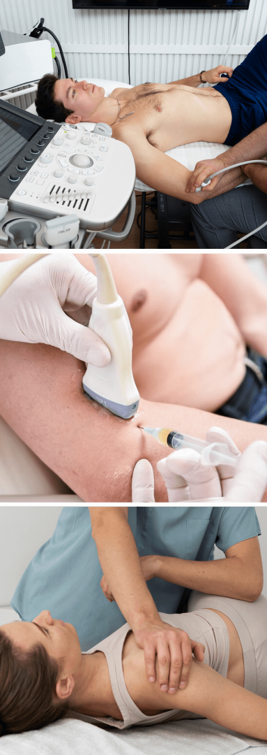
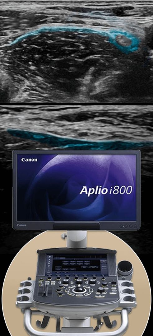
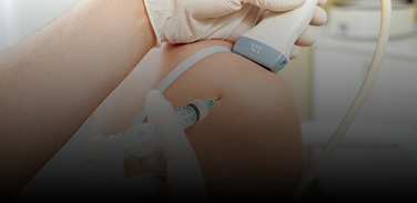
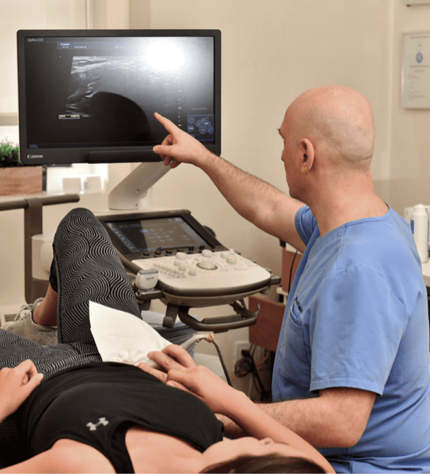
Unless your clinician is able to visualize your injured tissues and explore their impact on other structures, diagnosis is a guessing game at best. We use the highest-resolution diagnostic ultrasound to dynamically visualize your injury in real time.
Ultrasound imaging has several advantages over MRI:
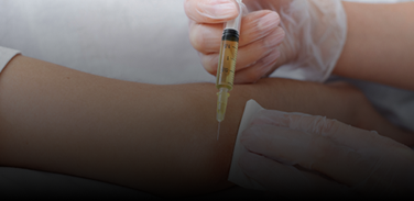
Once the diagnostic process is complete, we create a personalized treatment protocol, designed just for you. Your early treatment may include a combination of manual, regenerative and orthobiologic injection therapies aimed at healing damaged tissues prior to physical therapy.
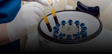
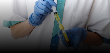
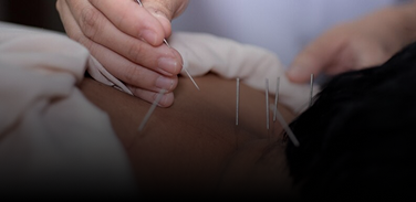
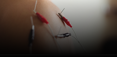
Orthobiologic injection therapies use natural/neutral solutions, injected with precision thanks to ultrasound guidance. The injected solutions stimulate cellular repair by either nourishing or irritating the targeted cells. Needling procedures like dry needling and PENS use filament-thin non-medicated needles to target myofascial trigger points and normalize neural activity.
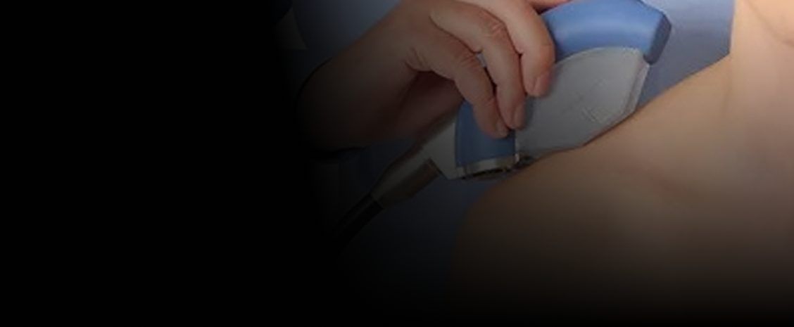
For ultrasound-guided needling procedures, Dr. Kalika partners with orthobiologic specialist Dr. Brosgol to ensure that the needles hit their mark. Treatment results are dramatically enhanced when combined with focused extracorporeal shockwave therapy (fESWT), and Stecco myofascial release, both areas of expertise for Dr. Kalika.
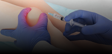
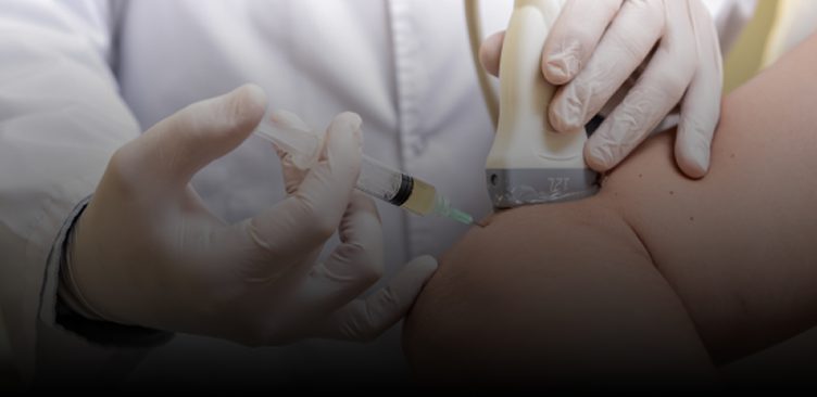
Without ultrasound imaging, therapeutic procedures are hit-or-miss, often failing to achieve their goals. Guidance by high resolution ultrasound ensures that injected solutions reach their intended tissues, without bleeding over into other structures. This means faster pain relief and accelerated healing, often with fewer treatment sessions.
During needling procedures, ultrasound guidance protects nerves and blood vessels from accidental needle penetration while ensuring that injected substances hit their target. It is important to note that only advanced high resolution ultrasound shows us minute details that cannot be seen with regular ultrasound imaging. High resolution imaging is critical for fascial and nerve injections, and for treating tendon tears.
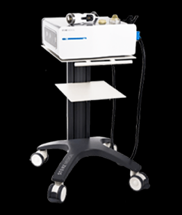
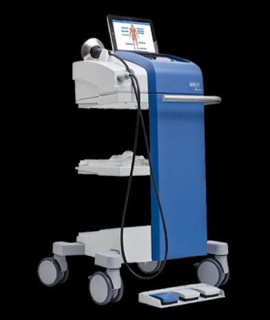
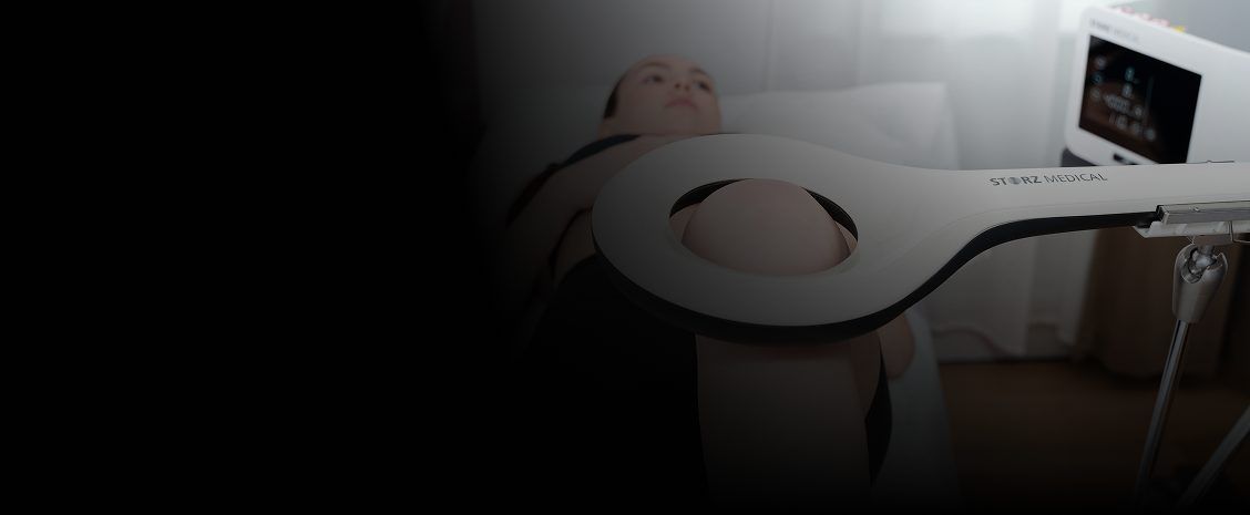
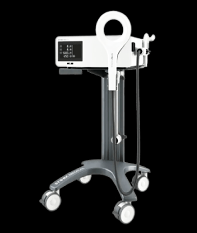
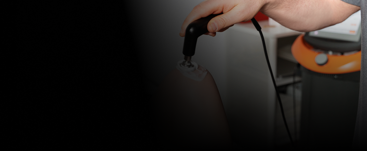
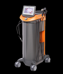
Regenerative technologies deliver natural energy waves – sound, radiofrequency, electric, and magnetic – to the injured area to stimulate and activate the body’s own innate healing mechanisms. These therapies are extracorporeal, meaning they are applied through the skin, without penetration.
When used in conjunction with injection therapies, regenerative technologies enhance and accelerate the healing effects of orthobiologics.
The success of regenerative and orthobiologic therapies requires extensive skills and knowledge on the part of the practitioner. Dr. Kalika’s expertise in human anatomy coupled with 20+ years of clinical experience have prepared him for these remarkable new approaches to integrative healing. His ongoing commitment to patient care and continuing education have placed him at the cutting edge of advanced healing methodologies.
Physical therapy is an important final step on your healing journey, but it should not be started prematurely. For physical therapy to provide effective and lasting results, we must first address structural issues and restore biotensegrity to the body’s systems. Advanced orthobiologic injection therapies play a critical role in injury rehab that cannot be filled by physical therapy alone. By the same token, injection therapies alone are not enough to complete your injury rehabilitation.
Once your pretreatment is complete, we can begin a customized physical therapy program with your specific goals in mind. Whether you want to return to sport as quickly as possible and perform at your peak, or you simply want to get back to your daily routine without pain or limitations, our personalized one-on-one approach ensures that your physical therapy prepares you for whatever lies ahead.
Muscles, fascia and nerves work interdependently to keep you on the move, and when one malfunctions, it affects the others. Healing damaged tissues and restoring biotensegrity are foundational to successful rehabilitation. Yet the vast majority of physical therapy clinics are poorly equipped in terms of knowledge, experience and technology.
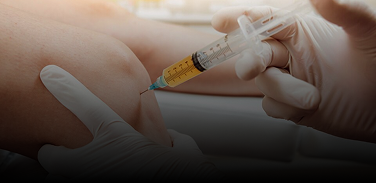
To be effective, injection therapies must precisely target tissues with the appropriate types and concentrations of injected solutions. “Blind” injections have a high potential of missing the targeted tissues altogether, and there is an elevated risk of penetrating other structures, like nerves and blood vessels.
At NYDNRehab, we guide our orthobiologic procedures with high resolution ultrasound, to ensure that the injected solution hits its mark without damaging other structures. The combined experience and expertise of doctors Kalika and Brosgol, along with our broad range of available therapies, make NYDNRehab the premier clinic for orthobiologic injection therapy and injury rehab in NYC.


Approaches like massage, foam rolling and other techniques to break up fascial adhesions and densifications – while they may provide some pain relief and temporarily improve mobility – do not activate the global nervous system. Stecco is a systematic approach that requires specific training. When done correctly, it not only provides short-term relief, but it sets in motion a cascade of reactions that reorganize collagen fibers, restore fascial gliding, reactivate neural connections, optimize muscle coordination patterns, and recalibrate motor control.


Independent peer-reviewed research relevant to this treatment approach.
Research authored or co-authored by the clinic’s medical director. The following research publications inform the clinical approach used in this treatment program.
Case report
2025
 Lev Kalika
  Bubnov Rostyslav
Below is a prime example of how ultrasound can take the guesswork out of diagnosis.

A bad physical therapy experience is one of the primary causes of unnecessary surgery
In this instance, an athlete was originally diagnosed with minor quadriceps muscle strain and was treated for four weeks, with unsatisfactory results. When he came to our clinic, the muscle was not healing, and the patients’ muscle tissue had already begun to atrophy.
Upon examination using MSUS, we discovered that he had a full muscle thickness tear that had been overlooked by his previous provider. To mitigate damage and promote healing, surgery should have been performed immediately after the injury occurred. Because of misdiagnosis and inappropriate treatment, the patient now has permanent damage that cannot be corrected.
The most important advantage of Ultrasound over MRI imaging is its ability to zero in on the symptomatic region and obtain imaging, with active participation and feedback from the patient. Using dynamic MSUS, we can see what happens when patients contract their muscles, something that cannot be done with MRI. From a diagnostic perspective, this interaction is invaluable.
Dynamic ultrasonography examination demonstrating
the full thickness tear and already occurring muscle atrophy
due to misdiagnosis and not referring the patient
to proper diagnostic workup
Demonstration of how very small muscle defect is made and revealed
to be a complete tear with muscle contraction
under diagnostic sonography (not possible with MRI)

Complete tear of rectus femoris
with large hematoma (blood)

Separation of muscle ends due to tear elicited
on dynamic sonography examination


