August 1, 2024
Musculoskeletal injuries from sports, trauma or overuse can be painful, messy and difficult to differentiate various layers and structures beneath the skin’s surface.
Ultrasound imaging can identify the exact location and nature of a musculoskeletal injury within seconds, enabling the health care provider to accurately diagnose and treat injured patients.
If you are not convinced that Ultrasonography is the best imaging technology for musculoskeletal injuries, here are 10 compelling reasons to become a believer.
While ultrasound technology is a relative late-comer to the diagnostic table, it is rapidly gaining ground as a superior form of imaging for musculoskeletal injuries.
For accurate diagnosis and treatment of musculoskeletal injuries and conditions, the sports medicine team at NYDNRehab relies on diagnostic ultrasonography and other technologies tom of your pain and heal it. Do not waste time on Xrays and MRI. Get accurate real-time imaging and treatment at NYDNRehab, so you can recover from your musculoskeletal injury and get back in the game.

Verified Expert Profiles
Dr. Lev Kalika is a world-recognized expert in musculoskeletal medicine. with 20+ years of clinical experience in diagnostic musculoskeletal ultrasonography, rehabilitative sports medicine and conservative orthopedics. In addition to operating his clinical practice in Manhattan, he regularly publishes peer-reviewed research on ultrasound-guided therapies and procedures. He serves as a peer reviewer for Springer Nature.
Dr. Kalika is an esteemed member of multiple professional organizations, including:
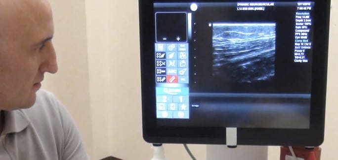
Below is a prime example of how ultrasound can take the guesswork out of diagnosis.

A bad physical therapy experience is one of the primary causes of unnecessary surgery
In this instance, an athlete was originally diagnosed with minor quadriceps muscle strain and was treated for four weeks, with unsatisfactory results. When he came to our clinic, the muscle was not healing, and the patients’ muscle tissue had already begun to atrophy.
Upon examination using MSUS, we discovered that he had a full muscle thickness tear that had been overlooked by his previous provider. To mitigate damage and promote healing, surgery should have been performed immediately after the injury occurred. Because of misdiagnosis and inappropriate treatment, the patient now has permanent damage that cannot be corrected.
The most important advantage of Ultrasound over MRI imaging is its ability to zero in on the symptomatic region and obtain imaging, with active participation and feedback from the patient. Using dynamic MSUS, we can see what happens when patients contract their muscles, something that cannot be done with MRI. From a diagnostic perspective, this interaction is invaluable.
Dynamic ultrasonography examination demonstrating
the full thickness tear and already occurring muscle atrophy
due to misdiagnosis and not referring the patient
to proper diagnostic workup
Demonstration of how very small muscle defect is made and revealed
to be a complete tear with muscle contraction
under diagnostic sonography (not possible with MRI)

Complete tear of rectus femoris
with large hematoma (blood)

Separation of muscle ends due to tear elicited
on dynamic sonography examination


