Most of us begin walking during our first year of life, toddling along until we get the hang of it, then developing the balance and coordination required to run, jump, skip and dance. But years of practice does not necessarily make perfect. Over time, we develop faulty movement patterns and imbalances that place wear and tear on our joints, muscles and tendons , and increases our risk of injury.
Gait analysis is not a new concept. Scientists have been using it for decades to study human function , improve athletic performance, rehabilitate injuries and promote functional motor skills in the elderly. But technology has given gait analysis a significant boost in recent years, changing our understanding of strength the way we analyze and retrain walking and running gait.
In recent decades, gait analysis results have relied primarily on two-dimensional videography to assess joint angles, with the technician drawing lines to indicate deficient motor patterns. The accuracy of 2D assessment relies largely on the skills and experience of the technician, and the perspective of the camera. Even at its best, 2D has a large margin of error, because it is limited to motion in the sagital plane of movement, where hip, knee and ankle flexion and extension take place.
However, the majority of walking and running injuries occur in the transverse plane of movement. If you had a camera looking straight down from above while you walked or ran, it would pick up rotational motion that cannot be detected, measured or evaluated with 2D. Without information about motion in the transverse plane, gait analysis has limited value for rehabilitation.
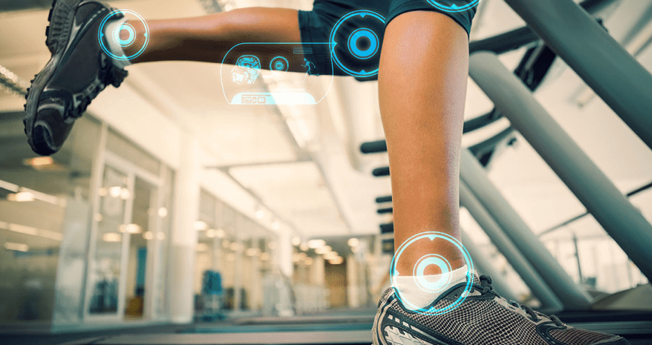
Today, the latest advances in technology have rendered 2D analysis almost obsolete. With 3D gait analysis, infrared cameras and reflective markers give a three dimensional view of walking and running gait.
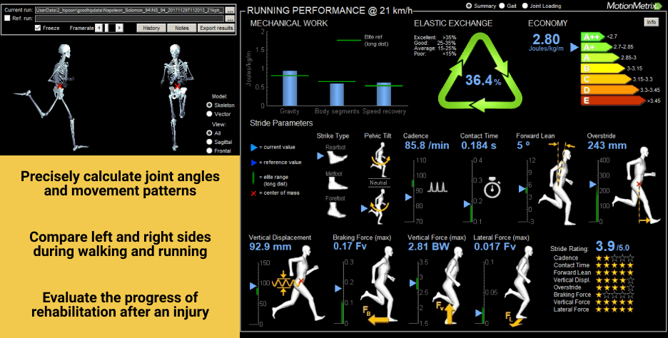
3D gait analysis can:
With data collected from 3D gait analysis, we can develop an individualized gait retraining program to improve gait, enhance performance and prevent injury.
All humans learn to walk and run at an early age, but imbalances can develop over time that reduce motor efficiency and increase the risk of injury. Whether you are an athlete wanting a competitive edge, recovering from an injury, need to improve your performance or simply want pain-free mobility for everyday function, gait analysis provides valuable data that helps us design a customized recovery program.
Pain syndromes and running performance are often caused by mechanical issues related to gait. Pain in your hip could be caused by faulty foot mechanics, or knee pain could be caused by imbalances in the pelvic region. Gait analysis helps us get to the source of the problem and correct it.
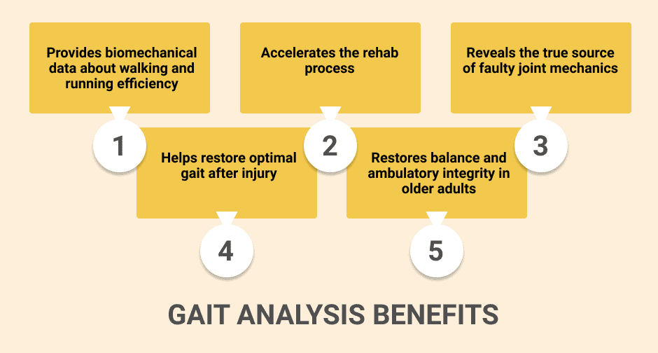
Gait analysis benefits include:
Gait analysis and retraining can help you enjoy a better quality of life on a daily basis, with less pain and reduced risk of injury from falls.
Whether you are an athlete in a sport that requires precise motor skills, a competitive runner looking for an edge or a novice athlete wanting to get started on the right foot, 3D gait analysis and retraining can be a complete game changer.
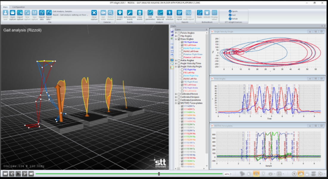
At NYDNRehab, we pride ourselves on keeping up to speed with the latest technologies and rehab approaches, to give our patients the best care possible. Our clinic is one of the few private clinics in the world equipped to give a comprehensive walking or running gait analysis using advanced technology. We possess motion analysis technologies beyond 3D gait analysis which allow us to zoom in into each lower extremity joints. The range of our technologies include all the tools used in modern academia ( just like the best university research labs in the country)to study human motion and performance.
Our gait analysis toolbox includes:
Don’t let faulty gait patterns keep you from doing the things you love. Contact NYDNRehab today, and schedule your 3D gait analysis for better performance, improved function and enhanced injury prevention.
Dr. Lev Kalika is clinical director of NYDNRehab, located in Manhattan. Lev Kalika is the author of multiple medical publications and research, and an international expert in the field of rehabilitative sonography, ultrasound guided dry needling and sports medicine Dr. Kalika works with athletes, runners, dancers and mainstream clients to relieve pain, rehabilitate injuries, enhance performance and minimize the risk of injuries. His clinic features some of the most technologically advanced equipment in the world, rarely found in a private clinic.


Brian Hanley, Catherine B. Tucker & Athanassios Bissas (2018) Differences between motion capture and video analysis systems in calculating knee angles in elite-standard race walking, Journal of Sports Sciences, 36:11, 1250-1255

Verified Expert Profiles
Dr. Lev Kalika is a world-recognized expert in musculoskeletal medicine. with 20+ years of clinical experience in diagnostic musculoskeletal ultrasonography, rehabilitative sports medicine and conservative orthopedics. In addition to operating his clinical practice in Manhattan, he regularly publishes peer-reviewed research on ultrasound-guided therapies and procedures. He serves as a peer reviewer for Springer Nature.
Dr. Kalika is an esteemed member of multiple professional organizations, including:
Below is a prime example of how ultrasound can take the guesswork out of diagnosis.

A bad physical therapy experience is one of the primary causes of unnecessary surgery
In this instance, an athlete was originally diagnosed with minor quadriceps muscle strain and was treated for four weeks, with unsatisfactory results. When he came to our clinic, the muscle was not healing, and the patients’ muscle tissue had already begun to atrophy.
Upon examination using MSUS, we discovered that he had a full muscle thickness tear that had been overlooked by his previous provider. To mitigate damage and promote healing, surgery should have been performed immediately after the injury occurred. Because of misdiagnosis and inappropriate treatment, the patient now has permanent damage that cannot be corrected.
The most important advantage of Ultrasound over MRI imaging is its ability to zero in on the symptomatic region and obtain imaging, with active participation and feedback from the patient. Using dynamic MSUS, we can see what happens when patients contract their muscles, something that cannot be done with MRI. From a diagnostic perspective, this interaction is invaluable.
Dynamic ultrasonography examination demonstrating
the full thickness tear and already occurring muscle atrophy
due to misdiagnosis and not referring the patient
to proper diagnostic workup
Demonstration of how very small muscle defect is made and revealed
to be a complete tear with muscle contraction
under diagnostic sonography (not possible with MRI)

Complete tear of rectus femoris
with large hematoma (blood)

Separation of muscle ends due to tear elicited
on dynamic sonography examination