January 22, 2025

Ankle sprains are extremely common among physically active populations, accounting for up to half of all sports injuries in the USA, and researchers estimate that fewer than half of ankle sprains sufferers seek medical attention for their injury. Of ankle injuries that do receive medical attention, as many as 40% are thought to be misdiagnosed or ineffectively treated, resulting in chronic ankle pain and instability.
Considering these facts, it is not only important to seek professional treatment for your ankle injury, but it is crucial to get an accurate diagnosis and personalized rehabilitation, to restore ankle function and reduce your risk of future injury.
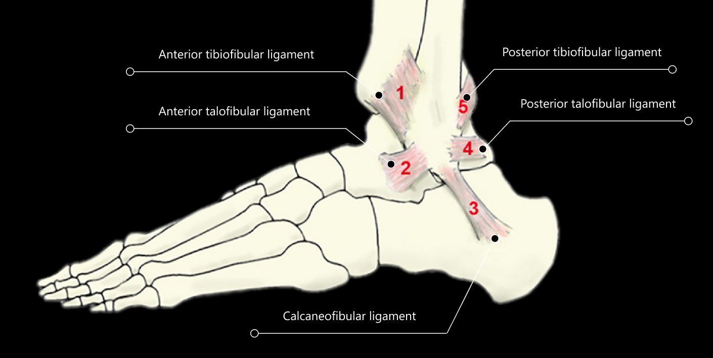
Ankle sprains involve stretching or tearing of ligaments – tough bands of connective tissue that support and stabilize the ankle bones. Ankle sprains range from mild to severe, depending on the number of ligaments involved and whether the ligament is merely stretched, partially torn, or completely torn.
Ankle sprains are different from ankle strains, which affect the soft tissues, or fractures that affect the bony structures. An ankle sprain can result in ankle instability due to ligament laxity that increases an athlete’s risk of injury – in fact, a previous sprain is the predominant risk factor for predicting a recurrent injury.
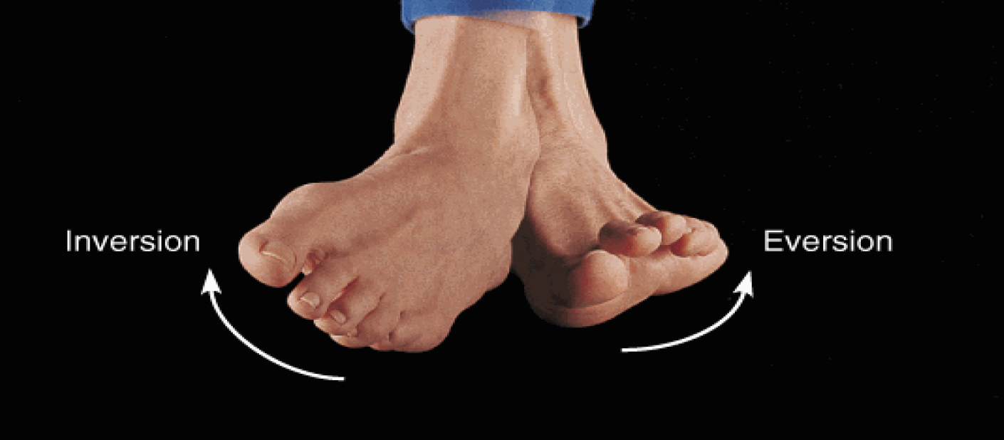
There are three basic categories of ankle sprains:
The severity of ankle sprains is graded on a scale of 1-3:

According to research, as many as 40% of ankle injuries are misdiagnosed or incorrectly treated. Other conditions that share similar symptoms are often mistaken for ankle sprains, especially when the diagnosis is symptoms-based. For example, high ankle sprains are often misdiagnosed as lateral sprains. Even imaging technologies like Xray or MRI can fail to adequately visualize the structures of the ankle, especially when inflammation is present.
Conditions that mimic ankle sprains include:
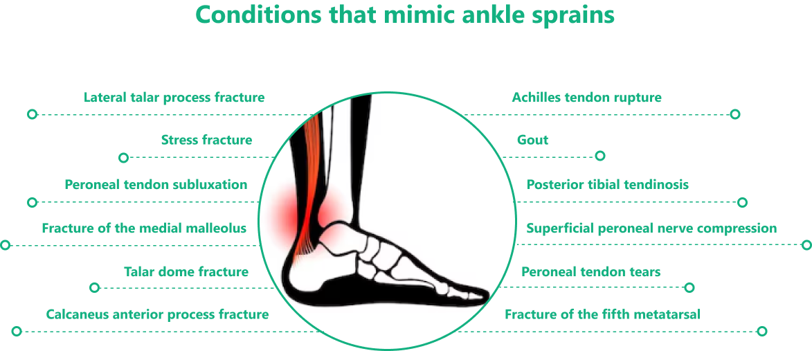
Misdiagnosis can lead to ineffective treatment that prolongs the patient’s pain and suffering while neglecting to adequately address damaged tissues. High-resolution diagnostic ultrasound imaging can help to accurately identify injured structures, and differentiate ankle sprains from other conditions.
Most physically active people are accustomed to various aches and pains from training and overuse, and many may ignore an ankle injury if it can still bear a load, even if it hurts. Over time, an untreated ankle sprain may stop hurting as inflammation goes down and tissues heal. But damaged tissues do not always heal effectively, and you may have scar tissue, ligament laxity, reduced range of motion, and reduced ankle stability, all of which can alter your gait, create compensation patterns, and increase your risk of recurrent injuries.
Here are 5 important reasons to seek holistic sprained ankle treatment and rehabilitation, even after you think your ankle has healed:
Ankle instability can affect your entire body due to reduced control of force loads. Structures that are not designed to manage loads may become strained, and compensation patterns may arise to help mediate forces during physical activity. Without rehabilitation, your ankle sprain can contribute to knee, hip, and back pain, and an untreated ankle injury can reduce your balance and increase your risk of falls as you age.
After an ankle sprain multiple factors can be affected by ankle instability, including:

One of the most common sensory deficits after an ankle sprain is the inability to estimate how much weight-bearing load your ankle can support. Sensory-motor function can be restored using a reweighing platform that provides perturbation, activating muscle receptors to help reprogram your brain-body connection for optimal motor control.
During sprained ankle rehabilitation, it is important to strengthen the ankle and its supporting structures, and to incorporate stretching exercises to regain full joint range of motion. But before loading the ankle, you may need to repair soft tissues that have healed poorly via treatments like shockwave therapy and orthobiologic injection therapy. Fascial densifications and adhesions may need to be addressed, to recover functional range of motion and restore muscle activation patterns.
Neglecting to rehabilitate your sprained ankle can lead to pain syndromes, poor balance and reduced mobility later in life. If you want to remain physically active and avoid future injuries, sprained ankle rehab is a must.
At NYDNRehab, we used advanced technologies to accurately diagnose and treat your ankle injury. Your healing journey begins with a comprehensive diagnostic exam using high-resolution ultrasonography. Dynamic ultrasound imaging lets us visualize your foot and ankle in real time, for quick and accurate diagnosis.
Our research-grade ultrasound equipment gives us capabilities for superior microvascular imaging (SMI) to detect early signs of blood flow to damaged tissues, and sonoelastography to test tissue density and elasticity.
Our human movement lab pairs state-of-the-art technology with proprietary software to give us quantitative information about various movement parameters, such as joint kinematics, load distribution, muscle coordination patterns and more. Our ultrasound-guided regenerative therapies and orthobiologic procedures help to restore tissue integrity, to optimize ankle function.
You don’t have to settle for low-tech, one-size-fits-all treatment protocols offered by conventional physical therapy clinics. Our personalized one-on-one approach to patient care means you will get customized treatment supported by advanced technologies and tailored to your specific needs. If you’re ready to rehab your sprained ankle and get moving again, contact NYDNRehab today.

Verified Expert Profiles
Dr. Lev Kalika is a world-recognized expert in musculoskeletal medicine. with 20+ years of clinical experience in diagnostic musculoskeletal ultrasonography, rehabilitative sports medicine and conservative orthopedics. In addition to operating his clinical practice in Manhattan, he regularly publishes peer-reviewed research on ultrasound-guided therapies and procedures. He serves as a peer reviewer for Springer Nature.
Dr. Kalika is an esteemed member of multiple professional organizations, including:
Below is a prime example of how ultrasound can take the guesswork out of diagnosis.

A bad physical therapy experience is one of the primary causes of unnecessary surgery
In this instance, an athlete was originally diagnosed with minor quadriceps muscle strain and was treated for four weeks, with unsatisfactory results. When he came to our clinic, the muscle was not healing, and the patients’ muscle tissue had already begun to atrophy.
Upon examination using MSUS, we discovered that he had a full muscle thickness tear that had been overlooked by his previous provider. To mitigate damage and promote healing, surgery should have been performed immediately after the injury occurred. Because of misdiagnosis and inappropriate treatment, the patient now has permanent damage that cannot be corrected.
The most important advantage of Ultrasound over MRI imaging is its ability to zero in on the symptomatic region and obtain imaging, with active participation and feedback from the patient. Using dynamic MSUS, we can see what happens when patients contract their muscles, something that cannot be done with MRI. From a diagnostic perspective, this interaction is invaluable.
Dynamic ultrasonography examination demonstrating
the full thickness tear and already occurring muscle atrophy
due to misdiagnosis and not referring the patient
to proper diagnostic workup
Demonstration of how very small muscle defect is made and revealed
to be a complete tear with muscle contraction
under diagnostic sonography (not possible with MRI)

Complete tear of rectus femoris
with large hematoma (blood)

Separation of muscle ends due to tear elicited
on dynamic sonography examination


