HomeBlogAbdominal Muscle Function After Diastasis Rectus Abominis (DRA)
Abdominal Muscle Function After Diastasis Rectus Abominis (DRA)
August 14, 2023
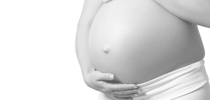
Diastasis rectus abominis (DRA) is a condition most commonly seen in post-partum females, although it occasionally occurs in obese individuals of both sexes. In a nutshell, DRA is a splitting of the linea alba, the thin but tough membrane that runs the length of the rectus abdominis (RA) muscle that defines the “six-pack.”
In some cases, DRA affects only a small section of the RA muscle, and there is little noticeable change in appearance or function. However, in severe cases, DRA can be both disfiguring and debilitating, compromising the function and performance of the RA and interfering with trunk and pelvic control.
RA Muscle Function
The RA muscle works to form a canister that protects the internal organs, stabilizes the spine and pelvis, assists in breathing expiration, and increases intra-abdominal pressure when sneezing, coughing, urinating, defecating, lifting and during childbirth.
With DRA, the function of the RA is partially or completely inhibited, causing a variety of symptoms, including:
• weak flabby abdomen • undesirable physical appearance • pain in low back, hip or pelvis • urinary and bowel problems • poor posture • feeling of weakness • pain during intercourse
Effect of Surgery on DRA
Considering that severe cases of DRA cannot self-heal, many sufferers seek surgical intervention tore function.
Conservative Treatment for DRA
Conservative treatment for DRA that focuses on retraining RA recruitment strategies has proven in many cases tore function, either partially or fully. Physical therapy for DRA may include the following:
Postural training: Because of the role the RA muscle plays in stabilizing your lower back and core, postural training focuses on learning to perform daily activities like lifting your baby while maintaining proper posture.
Stretching: When muscles in one area of you body become weak, you may compensate by overusing other muscles, resulting in tightness that interferes with performance. Your therapist may prescribe stretching exercises tore balanced muscle tension.
Bracing: Your therapist may recommend taping or bracing your low back and abdominal region to decreases pain.
Education: Your therapist will help you understand which movements tore full function.
Specialized DRA Treatment Therapies at NYDNRehab
The physical therapy team at NYDNRehab uses the most innovative and effective therapies currently available for DRA:
Vojta Therapy works by reflex activation of the deep abdominal muscles, “reminding” your nervous system of how to effectively recruit them.
DNS (dynamic neuromuscular stabilization) and ISM (integrated system model) therapies take deep abdominal training to the next level, increasing strength and stability and promoting structural balance.
Failure to recovery from DRA.
Follow me:
About the Author
Dr. Lev Kalika is a world-recognized expert in musculoskeletal ultrasonography, with 20+ years of clinical experience in advanced rehabilitative medicine. In addition to operating his clinical practice in Manhattan, he regularly publishes peer-reviewed research on ultrasound-guided therapies and procedures.
Dr. Kalika is an esteemed member of the International Society for Medical Shockwave Treatment ((SMST), and the only clinician in New York certified by the ISMST to perform extracorporeal shockwave therapy. He is also an active member of the American Institute of Ultrasound in Medicine (AIUM), and has developed his own unique approach to dynamic functional and fascial ultrasonography.
In this instance, an athlete was originally diagnosed with minor quadriceps muscle strain and was treated for four weeks, with unsatisfactory results. When he came to our clinic, the muscle was not healing, and the patients’ muscle tissue had already begun to atrophy.
Upon examination using MSUS, we discovered that he had a full muscle thickness tear that had been overlooked by his previous provider. To mitigate damage and promote healing, surgery should have been performed immediately after the injury occurred. Because of misdiagnosis and inappropriate treatment, the patient now has permanent damage that cannot be corrected.
The most important advantage of Ultrasound over MRI imaging is its ability to zero in on the symptomatic region and obtain imaging, with active participation and feedback from the patient. Using dynamic MSUS, we can see what happens when patients contract their muscles, something that cannot be done with MRI. From a diagnostic perspective, this interaction is invaluable.
Dynamic ultrasonography examination demonstrating the full thickness tear and already occurring muscle atrophy due to misdiagnosis and not referring the patient to proper diagnostic workup
Demonstration of how very small muscle defect is made and revealed to be a complete tear with muscle contraction under diagnostic sonography (not possible with MRI)
Complete tear of rectus femoris with large hematoma (blood)
Separation of muscle ends due to tear elicited on dynamic sonography examination