Your hamstrings work together with your gluteal muscles to extend your hip during physical activity, and while your glutes do the lion’s share of the work, sometimes your hamstrings succumb to overuse, especially if they are not adequately trained.
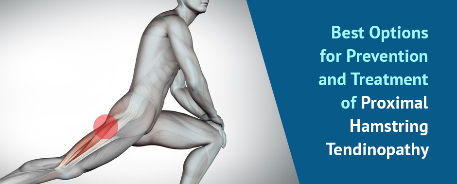
If you are experiencing hamstring and buttock pain, you may have a condition called proximal hamstring tendinopathy (PHT). Learn more about the condition, and the best and fastest available treatment options.
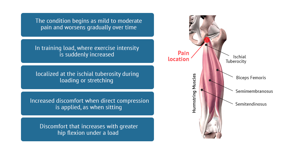
There are four muscles at the back of each of your thighs that make up the hamstrings. Your hamstrings cross over two joints — at the knee where they produce knee flexion, and at the hip, where they contribute to hip extension. The proximal tendons of three of the hamstring muscles are anchored to the pelvic ischial tuberosity, the lower part of the pelvis beneath the buttocks, sometimes called the “sit bones.”
Repetitive use during sports or exercise can cause the hamstring tendons to become inflamed and irritated, resulting in pain and reduced mobility.In severe cases, the tendon may even become torn or ruptured. Excessive sitting can also cause upper hamstring pain in people who are sedentary and/or elderly.
Over time, if left untreated, the tendon may begin to degenerate, a condition called proximal hamstring tendinopathy that impedes function and reduces athletic performance.
A recent study interviewed 13 expert physical therapists and analyzed data to gather information on the diagnosis, management and prevention of PHT.
According to the experts, characteristics of the condition include:
An insidious onset, meaning the condition begins as mild to moderate pain and worsens gradually over time
Pain following a spike in training load, where exercise intensity is suddenly increased, such as when a runner increases their distance before an event, or when hill running is added to their training regimen
Pain that is consistently localized at the ischial tuberosity during loading or stretching, and does not radiate to other parts of the body
Increased discomfort when direct compression is applied, as when sitting
Discomfort that increases with greater hip flexion under a load
During diagnosis, the clinician will seek to differentiate your condition from other possible causes of pain, to determine if you have simple tendinitis, a tendon rupture or avulsion, a compressed or entrapped sciatic nerve, or proximal hamstring tendinopathy.
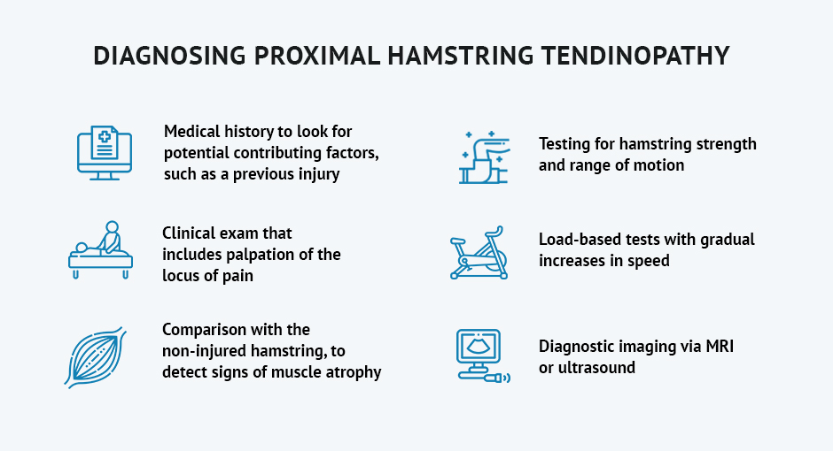
Diagnosis may include:
Medical history to look for potential contributing factors, such as a previous injury
Clinical exam that includes palpation of the locus of pain
Comparison with the non-injured hamstring, to detect signs of muscle atrophy
Testing for hamstring strength and range of motion
Load-based tests with gradual increases in speed
Diagnostic imaging via MRI or ultrasound
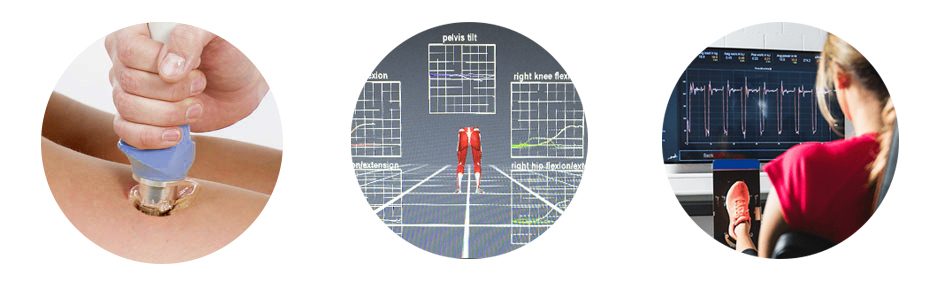
High resolution diagnostic ultrasound is the imaging method of choice for upper hamstring pain, because it allows us to visualize the injured area in real time, with the patient in motion. It also enables us to scan the entire length of structures like bones and nerves, to look for issues like sciatic nerve pain in the hamstrings.
The experts agree that there is no quick fix for PHT, which can be frustrating for athletes and fitness enthusiasts who want to remain active. Reducing physical activity and avoiding exercises or activities that involve even moderate hip flexion is necessary during the rehab and healing process.
Treatment options for PHT include:
Manual hamstring massage
Blood flow restriction training
Progressive eccentric loading
Biomechanical analysis and retraining
Gait analysis and retraining
Working with a physical therapist who specializes in tendon issues can mean the difference between full recovery from proximal hamstring tendinopathy and return to normal physical activity, or resigning yourself to reduced activity and potential long-term disability.
Tendon injuries are common among physically active adults, and neglecting to treat them can lead to tendon degeneration, reduced function and limited mobility. Even if you have lived with tendinopathy for years after a tendon injury, there is still a chance for full recovery.
The tendon specialists at NYDNRehab used the latest therapies and most advanced technologies to rehabilitate tendon injuries. Our clinic features the highest resolution diagnostic ultrasound equipment available, for fast and accurate diagnosis. Dr. Kalika is a certified expert in diagnostic ultrasonography, ultrasound guided dry needling, ESWT and other regenerative therapies that stimulate healing at the cellular level.
If you want to eliminate upper hamstring pain and get back to your favorite sport or physical activity, contact NYDNRehab today, and jump-start the healing process so you can get back to doing the things you love!
Resource
Nasser, Anthony M., et al. “Proximal hamstring tendinopathy; expert physiotherapists’ perspectives on diagnosis, management and prevention.” Physical Therapy in Sport 48 (2021): 67-75.

Verified Expert Profiles
Dr. Lev Kalika is a world-recognized expert in musculoskeletal medicine. with 20+ years of clinical experience in diagnostic musculoskeletal ultrasonography, rehabilitative sports medicine and conservative orthopedics. In addition to operating his clinical practice in Manhattan, he regularly publishes peer-reviewed research on ultrasound-guided therapies and procedures. He serves as a peer reviewer for Springer Nature.
Dr. Kalika is an esteemed member of multiple professional organizations, including:
Below is a prime example of how ultrasound can take the guesswork out of diagnosis.

A bad physical therapy experience is one of the primary causes of unnecessary surgery
In this instance, an athlete was originally diagnosed with minor quadriceps muscle strain and was treated for four weeks, with unsatisfactory results. When he came to our clinic, the muscle was not healing, and the patients’ muscle tissue had already begun to atrophy.
Upon examination using MSUS, we discovered that he had a full muscle thickness tear that had been overlooked by his previous provider. To mitigate damage and promote healing, surgery should have been performed immediately after the injury occurred. Because of misdiagnosis and inappropriate treatment, the patient now has permanent damage that cannot be corrected.
The most important advantage of Ultrasound over MRI imaging is its ability to zero in on the symptomatic region and obtain imaging, with active participation and feedback from the patient. Using dynamic MSUS, we can see what happens when patients contract their muscles, something that cannot be done with MRI. From a diagnostic perspective, this interaction is invaluable.
Dynamic ultrasonography examination demonstrating
the full thickness tear and already occurring muscle atrophy
due to misdiagnosis and not referring the patient
to proper diagnostic workup
Demonstration of how very small muscle defect is made and revealed
to be a complete tear with muscle contraction
under diagnostic sonography (not possible with MRI)

Complete tear of rectus femoris
with large hematoma (blood)

Separation of muscle ends due to tear elicited
on dynamic sonography examination


