
For an injured athlete, the rehabilitation process can seem slow and arduous. Time taken away from sport and training can be costly on many levels, reducing opportunities for advancement and reversing hard-earned performance gains. In the world of sports, innovative therapies that speed up healing and accelerate rehab time are in constant demand. One such innovation is blood flow restriction therapy, or BFRT.
While BFRT is a relatively new approach for injury rehab, the method has been used since the 1960s as a training tool, enabling athletes to build lean muscle mass without excessive weight loading. BFRT originated in Japan as KAATSU training in 1966. The idea is to apply pressure to an arm or leg during exercise to reduce venous blood flow from the limb while allowing normal arterial blood to flow into the muscle.
Prior to being used for injury rehab, BFRT was used by the military and by professional sports teams as a training aid. Years of research and multiple upgrades to the approach have expanded the popularity of BFRT, and gained the attention of the rehabilitation community.
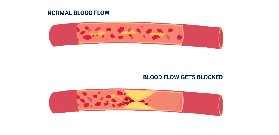
The best way to visualize BFRT is to imagine an inflated blood pressure cuff applied to your upper arm or thigh prior to beginning an exercise set. As arterial blood flows into the muscles, venous blood is retained, causing the blood to pool. The pooling of blood brings on volitional fatigue at much lower training loads than conventional resistance exercise, eliciting an increase in strength and size at a much lower training volume.
Conventional resistance training loads are calculated at about 70% of one-repetition maximum (1RM), the total amount the individual can lift only once. A conventional protocol for muscle hypertrophy calls for volitional fatigue somewhere between 6 to 10 repetitions, repeated for 3 or more sets. With BFRT, loading is calculated at 20 to 30% of 1RM, with 15 to 30 repetitions per set, for 3 to 5 sets. Lower weights with higher reps and more sets is the accepted BFRT formula for optimal results.
Although the exact mechanisms for muscle adaptation are unclear, it is suspected that BFRT stimulates certain chemical reactions that normally occur with higher training loads, enhancing muscle strength and triggering muscle hypertrophy without heavy loading.

Because BFRT uses lower training loads than conventional resistance training, it reduces strain on injured joints, bones and soft tissues while enabling the patient to build strength and increase muscle mass during recovery. For example, a football player with a ruptured ACL can train the quadriceps muscles without overloading the knee joint, or a swimmer with a torn rotator cuff can train the biceps without overloading the shoulder.
Individuals who can benefit from BFRT include:
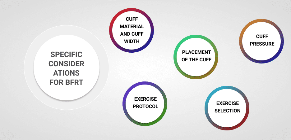
As BFRT has grown in popularity, the procedures for its application have become more refined. A specialist trained in BFRT understands the nuances of the approach, and is able to get the best results.
Specific considerations for BFRT include:
In response to popular demand, specialized equipment and training have emerged to ensure the best results from BFRT. To get the most from a BFRT training session, find a specialist who has been trained to use the latest BFRT equipment, to get the best results. BFRT should be combined with other rehab methods to ensure a well-rounded and complete recovery protocol.
NYDNRehab is a premier rehab clinic in NYC that outpaces other clinics with advanced technologies and innovative treatment methods. Blood flow restriction therapy is one of the many tools we use to rehabilitate injured athletes and other patients who can benefit from low-load training.
If you are injured and want to recover as quickly as possible, contact NYDNRehab. Our specialists will design a customized program based on your particular needs and circumstances. We are dedicated to getting you back to the things you love, with improved performance and reduced risk of re-injury.

Verified Expert Profiles
Dr. Lev Kalika is a world-recognized expert in musculoskeletal medicine. with 20+ years of clinical experience in diagnostic musculoskeletal ultrasonography, rehabilitative sports medicine and conservative orthopedics. In addition to operating his clinical practice in Manhattan, he regularly publishes peer-reviewed research on ultrasound-guided therapies and procedures. He serves as a peer reviewer for Springer Nature.
Dr. Kalika is an esteemed member of multiple professional organizations, including:
Below is a prime example of how ultrasound can take the guesswork out of diagnosis.

A bad physical therapy experience is one of the primary causes of unnecessary surgery
In this instance, an athlete was originally diagnosed with minor quadriceps muscle strain and was treated for four weeks, with unsatisfactory results. When he came to our clinic, the muscle was not healing, and the patients’ muscle tissue had already begun to atrophy.
Upon examination using MSUS, we discovered that he had a full muscle thickness tear that had been overlooked by his previous provider. To mitigate damage and promote healing, surgery should have been performed immediately after the injury occurred. Because of misdiagnosis and inappropriate treatment, the patient now has permanent damage that cannot be corrected.
The most important advantage of Ultrasound over MRI imaging is its ability to zero in on the symptomatic region and obtain imaging, with active participation and feedback from the patient. Using dynamic MSUS, we can see what happens when patients contract their muscles, something that cannot be done with MRI. From a diagnostic perspective, this interaction is invaluable.
Dynamic ultrasonography examination demonstrating
the full thickness tear and already occurring muscle atrophy
due to misdiagnosis and not referring the patient
to proper diagnostic workup
Demonstration of how very small muscle defect is made and revealed
to be a complete tear with muscle contraction
under diagnostic sonography (not possible with MRI)

Complete tear of rectus femoris
with large hematoma (blood)

Separation of muscle ends due to tear elicited
on dynamic sonography examination


