February 1, 2024
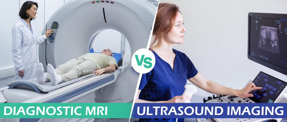 Diagnostic imaging modalities like X-ray, CT scan and MRI represented a quantum leap forward in 20th Century medicine, enabling doctors to look beneath the skin to visualize bones, joints, and soft tissues. But those breakthrough technologies have certain limitations in terms of risk, convenience, cost and utility.
In the 21st Century, ultrasonography has taken diagnostic imaging to the next level, providing a safer, faster and more efficient mode of visualizing the body’s structures.
Learn the fundamentals of MRI vs ultrasound technology, and their advantages and disadvantages for diagnosing disease, metabolic disorders, and musculoskeletal injuries, pain and dysfunction.
Magnetic resonance imaging (MR!) technology took the lead in the 1980s as an advanced diagnostic tool, enabling clinicians to visualize the body’s anatomy without the damaging ionizing radiation of X Rays and CT scans. In the years to follow, MRI became the gold standard for medical diagnostic imaging.
MRI uses powerful magnets to produce a strong magnetic field, tapping into the body’s innate magnetic properties to produce detailed images of the body’s organs and structures. The magnetic field causes the body’s atoms to align in the same direction. Radio waves are then sent from the MRI machine, moving the atoms out of their original position. As the radio waves are turned off, the atoms realign with the magnetic field, and MRI sensors are able to produce images based on the energy released. Clinicians can then analyze the images on a monitor to distinguish between different types of tissues — bones, muscles, nerves —based on their magnetic properties.

The discovery of magnetic resonance technology is credited to Austrian-born physicist Isidor Isaac Rabi whose family moved to the US in 1899. In the 1930s, Rabi began experimenting with magnetic fields and the nuclear spin of atoms. His research soon led to the magnetic resonance method, forming the basis for magnetic resonance imaging.
After World War II, other scientists began experimenting with the magnetic properties of atoms and molecules, further developing the imaging technique used for medical diagnosis. The first live images of a human subject were produced in 1977, and MRI machines became commercially available in the 1980s.

In the latter half of the 20th Century, ultrasound technology arrived on the scene. Ultrasonography uses high-frequency sound waves to produce dynamic images of organs, tissues, nerves and blood vessels. When directed at internal structures, the sound waves echo back, creating images that represent the size, shape and actions of the targeted tissues.
Ultrasound technology was originally developed as an industrial tool to detect flaws in welding. A Scottish obstetrician diverted the technology to investigate whether it could differentiate between soft tissue samples, and his early research was published in The Lancet in 1958.
As a radiation-free technology, ultrasonography was quickly adopted to view human fetuses in utero. The world’s first commercial ultrasound scanner became available in 1963. and the first obstetric ultrasounds were performed in the 1960s, with images of the placenta and a pulsing fetal heartbeat visible at nine weeks.
Despite the technology’s success, early ultrasound images were fuzzy and difficult to interpret, limiting its use as a diagnostic tool. But advancements in technology over the next several decades enabled enhanced imaging of the body’s structures, expanding its use from obstetrics to a broad range of other medical disciplines.

Today, the resolution and clarity of ultrasound images rival those of MRI, making ultrasonography a top contender as the diagnostic imaging modality of choice in the 2020s.
Magnetic resonance imaging is conducted without ionized radiation, making it a safer choice than Xray or CT scan. But the pulsed gradient magnetic field generated by MRI and its radiofrequency energy still pose certain risks.
The strong magnetic field produced by the MRI scanner pulls on magnetic materials, making it a high-risk diagnostic option for patients who use internal or external medical devices. MRI affects implanted devices like artificial joints, stents, cochlear implants, and pacemakers. It can also affect external devices like insulin pumps, metallic braces, and wound dressings.
Changes in MRI’s magnetic field and radio frequencies can cause devices and tissues to heat up, resulting in burns. According to the FDA, second degree burns are the most commonly reported side effect of MRI. The technology can also cause electronic devices like pacemakers to malfunction. At the same time, the presence of a medical device can degrade the quality of MRI images.
In addition to issues related to MRI magnetic and energy fields, gadolinium-based contrast agents (GBCAs) are often injected into the patient’s bloodstream via IV to enhance the visualization of internal organs, blood vessels and tissues. In 2018, the FDA released a warning about GBCAs used with MRI.
GBCAs contain heavy metals that must be eliminated by the kidneys, making MRI a poor choice for patients with kidney problems. Even in patients with healthy kidneys, heavy metals may remain in the body for months or even years, and may cross over into the brain. Gadolinium toxicity can produce a variety of unpleasant side effects ranging from mild to severe.
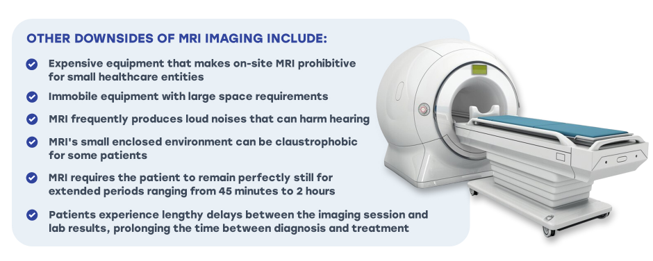
Other downsides of MRI imaging include:
Certain people should avoid MRI, including those with:
Diagnostic ultrasonography has multiple advantages over MRI. High-resolution ultrasound produces crystal-clear images that are easy to interpret, without the use of radiation or toxic heavy metals. Patient discomfort is minimal, and results are instantaneous, expediting appropriate treatment.
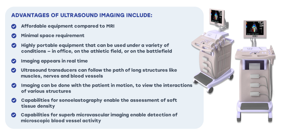
Advantages of ultrasound imaging include:
 Dynamic Ultrasonography for Next-Level Imaging of Sports Injuries
Dynamic ultrasound using the UNOSO ProbeFix device lets clinicians visualize damaged tissues during sport-specific actions, rendering fast, accurate real-time images of the body’s structures in motion. The device can even be synced with motion capture cameras to render 3D images of muscles, fascia, bones and joints during physical activity.
Capabilities of dynamic ultrasound imaging include:
Dynamic ultrasonography is a game changer for high-performance individuals who want to perfect dynamic movement and fine-tune sport-specific skills based on biomechanical efficiency. The ability to participate in the imaging process and watch the images in real time includes the patient as a key stakeholder in their diagnosis and treatment.
Your body is designed to move, and your body’s structures are meant to be interactive and interdependent. High resolution ultrasound imaging gives us a cutting-edge tool for visualizing your bones, muscles, organs and tissues in motion, leading to a more precise and accurate diagnosis than MRI.
The clinic at NYDNRehab features the most advanced musculoskeletal ultrasound equipment available. We are one of the first private clinics in NYC to leverage dynamic UNOSO technology. Dr. Kalika’s training and experience in diagnostic ultrasonography has positioned him as a recognized expert in diagnostic ultrasonography, with multiple scientific publications to his credit.
To get the most accurate diagnosis possible, followed by fast and effective treatment, contact NYDNRehab today!

Verified Expert Profiles
Dr. Lev Kalika is a world-recognized expert in musculoskeletal medicine. with 20+ years of clinical experience in diagnostic musculoskeletal ultrasonography, rehabilitative sports medicine and conservative orthopedics. In addition to operating his clinical practice in Manhattan, he regularly publishes peer-reviewed research on ultrasound-guided therapies and procedures. He serves as a peer reviewer for Springer Nature.
Dr. Kalika is an esteemed member of multiple professional organizations, including:
Below is a prime example of how ultrasound can take the guesswork out of diagnosis.

A bad physical therapy experience is one of the primary causes of unnecessary surgery
In this instance, an athlete was originally diagnosed with minor quadriceps muscle strain and was treated for four weeks, with unsatisfactory results. When he came to our clinic, the muscle was not healing, and the patients’ muscle tissue had already begun to atrophy.
Upon examination using MSUS, we discovered that he had a full muscle thickness tear that had been overlooked by his previous provider. To mitigate damage and promote healing, surgery should have been performed immediately after the injury occurred. Because of misdiagnosis and inappropriate treatment, the patient now has permanent damage that cannot be corrected.
The most important advantage of Ultrasound over MRI imaging is its ability to zero in on the symptomatic region and obtain imaging, with active participation and feedback from the patient. Using dynamic MSUS, we can see what happens when patients contract their muscles, something that cannot be done with MRI. From a diagnostic perspective, this interaction is invaluable.
Dynamic ultrasonography examination demonstrating
the full thickness tear and already occurring muscle atrophy
due to misdiagnosis and not referring the patient
to proper diagnostic workup
Demonstration of how very small muscle defect is made and revealed
to be a complete tear with muscle contraction
under diagnostic sonography (not possible with MRI)

Complete tear of rectus femoris
with large hematoma (blood)

Separation of muscle ends due to tear elicited
on dynamic sonography examination


