December 27, 2024

The kinetic chain (aka kinetic link) concept describes human movement in terms of a series of interrelated segments, and how movement of one link in the chain affects other segments. The kinetic chain principle gives us a framework for understanding human movement patterns in terms of muscle action, joint alignment, exercise conditioning and injury rehabilitation.
Your kinetic chain has five key points that link its individual segments:
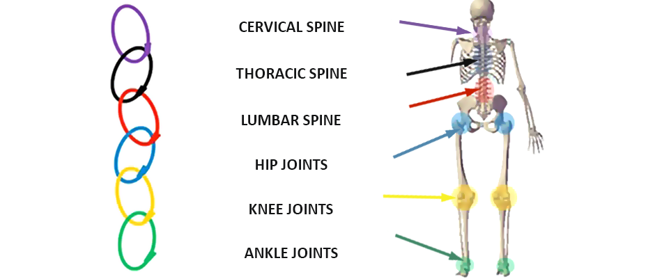
To better understand the concept, imagine an actual metal chain. You may have heard the adage, “a chain is only as strong as its weakest link.” When forces are applied to either end of the chain, a weakened link reduces the ability of the entire chain to to withstand applied forces. By analogy, malfunction of one or more links in the human kinetic chain can compromise the integrity of other links anywhere along the chain.
For example, if you sprain your ankle, your brain signals your muscles to compensate by shifting weight onto the uninjured limb. Not only does this affect the knee, hip and pelvis on the injured side, but on the uninjured side as well, as those segments bear greater-than-normal loads. Over time, prolonged compensation patterns rewire the body’s neural pathways, adapting muscle firing patterns to accommodate modifications in load distribution.
When it comes to rehabilitation of an ankle injury, it is not enough to treat the site of injury. We also need to strengthen structures along the kinetic chain and restore optimal muscle coordination, to redistribute force loads and eliminate compensation patterns. Failure to fully rehabilitate related structures and restore muscle firing patterns can cause more problems down the road and increase your risk of re-injury.
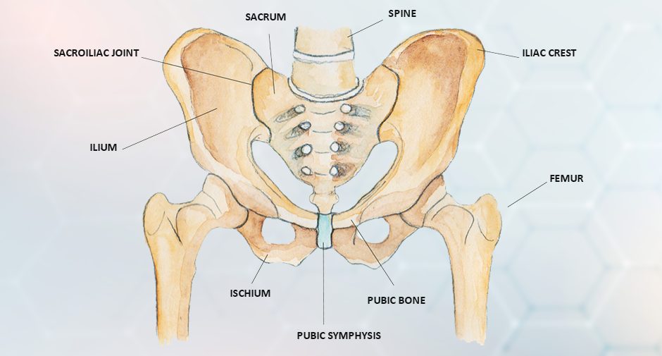
As the body’s centerpoint, the pelvic region is a pivotal link in your kinetic chain. A strong and stable pelvis governs the alignment of body segments both upward and downward along the chain. The bony structures of the pelvis and the pelvic floor muscles make up what is called the “pelvic bowl.” The pelvic bowl concept is useful in understanding pelvic alignment.
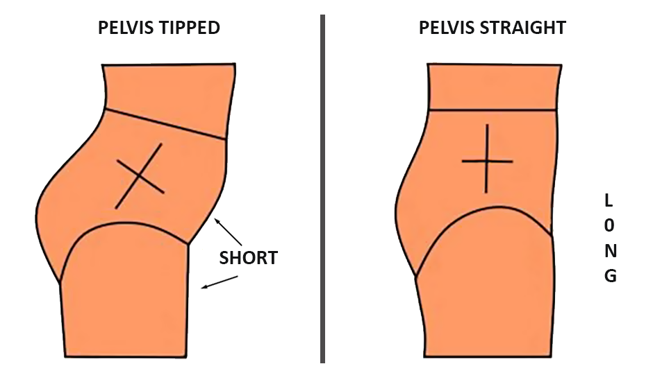
Imagine filling your pelvic bowl with water. When the pelvis is optimally aligned, the water is level and stays inside the bowl — this is called neutral pelvic alignment.
If you tilt the bowl backward, water spills out the back, and you are said to have a posterior pelvic tilt. Likewise, if you tilt the bowl forward, water spills out the front due to an anterior (frontward) pelvic tilt. Whether tilted forward or backward, your pelvis becomes misaligned, and that misalignment translates upward and downward to other segments along your kinetic chain.
Good pelvic alignment relies on a balance of forces generated by the muscles that come together at the pelvis and attach to the bones of the pelvic bowl. They include the gluteal and hamstring muscles in the back, and the hip flexors and abdominal muscles in the front. Your spinal extensors and pelvic floor muscles also play a role. When one or more of those muscle groups become too tight or too lax, it can force your pelvis out of alignment.
Key contributing factors that cause pelvic misalignment are physical inactivity and too much sitting. Your body is designed to walk, run and jump during waking hours, recruiting muscle groups in coordinated patterns that promote balanced muscle tension. Sitting on a chair for hours on end, day after day, is the fastest way to develop an anterior pelvic tilt.
On the other hand, people who stand on their feet for long hours every day often develop postural habits that cause the pelvis to tilt backward. Behaviors like locking the knees, shifting the body weight to one leg, disengaging the abdominal muscles, clenching the glutes and resting the weight of the upper body on the pelvis can cause posterior pelvic tilt.
In primitive cultures, minimal time was spent sitting, and even then people either squatted or sat cross-legged on the ground. In those positions, the spine remains neutral, the abdominal muscles engage to maintain stability, and the pelvic floor relaxes.
By contrast, when seated on a chair, the abdominal muscles remain lax while the spinal extensors are engaged. The hip flexors are shortened, the gluteal muscles lengthened, and the pelvis tilts forward, placing pressure on the pelvic floor. At the same time, the upper spine and shoulders often round forward, with the head jutting forward from its natural alignment.
Over time, weakened abdominal muscles and lax glutes, combined with shortened hip flexors and tight spinal extensors, cause the pelvic to tilt forward when sitting, standing or walking. This causes the lumbar spine to curve inward, compressing the space between the lumbar vertebrae and placing pressure on the intervertebral discs — a posture called lordosis.
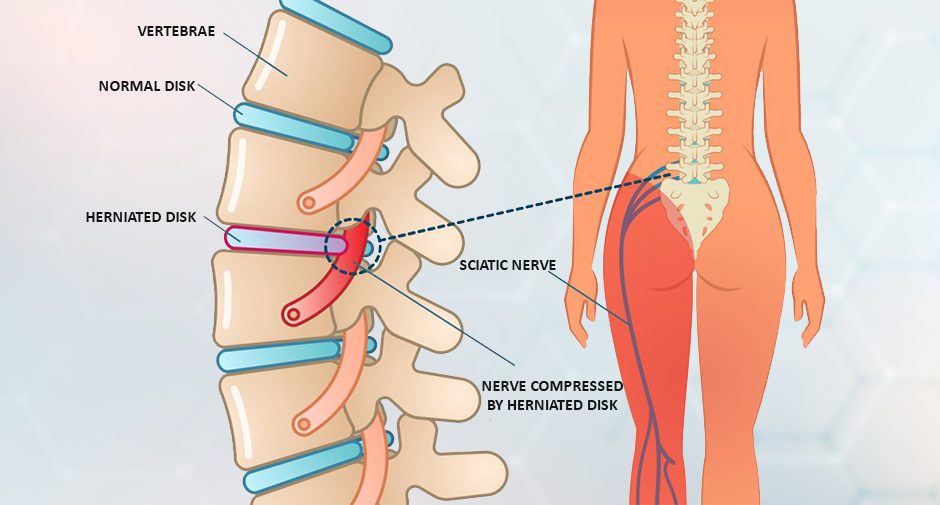
Intervertebral compression can cause the discs to bulge or herniate, irritating the nerve roots and causing pain. When the space between L4 and L5 is compressed, it can trigger sciatica, causing pain that travels from the low back to the buttocks and down the back of the leg.
Both anterior and posterior pelvic tilt can cause intervertebral compression and low back pain because they force the lumbar spine out of its neutral alignment. Pelvic misalignment can also cause nerve compression and pain anywhere along the spinal kinetic chain, all the way to the neck and head.
To reclaim good pelvic alignment, you need to address muscle imbalances. Depending on your current physical condition, this can be a quick fix, or it could take several months. Confounding factors are excess body weight, harmful lifestyle behaviors, metabolic disorders, and constraints imposed by occupation.
A good place to start is daily walking. The human body is well designed to walk and carry objects. Devoting 30-60 minutes a day to walking – in either a single bout or in shorter spurts – will automatically lengthen your hip flexors, strengthen your gluteal and hamstring muscles, engage your abdominal and pelvic floor muscles, and neutralize your spine.
To get the most out of walking:
If walking is painful or uncomfortable for you, consider getting a comprehensive gait analysis to pinpoint and correct mechanical issues.
Adding whole-body activities like yoga and resistance training can take you to the next level, enhancing your mobility and stability, and helping you to reach and maintain a healthy body weight. To put the icing on the cake, consider a biomechanical analysis followed by corrective physical therapy to help you achieve peak physical performance and pain-free mobility.
The physical therapy team at NYDNRehab is ready to help you get in the best shape of your life by correcting pelvic tilt, optimizing body alignment, improving posture and balancing muscle tension.
Don’t wait until back pain sets in to fix your pelvic tilt. Contact NYDNRehab today, and learn how to improve your health by balancing your muscles and realigning your kinetic chain.

Verified Expert Profiles
Dr. Lev Kalika is a world-recognized expert in musculoskeletal medicine. with 20+ years of clinical experience in diagnostic musculoskeletal ultrasonography, rehabilitative sports medicine and conservative orthopedics. In addition to operating his clinical practice in Manhattan, he regularly publishes peer-reviewed research on ultrasound-guided therapies and procedures. He serves as a peer reviewer for Springer Nature.
Dr. Kalika is an esteemed member of multiple professional organizations, including:
Below is a prime example of how ultrasound can take the guesswork out of diagnosis.

A bad physical therapy experience is one of the primary causes of unnecessary surgery
In this instance, an athlete was originally diagnosed with minor quadriceps muscle strain and was treated for four weeks, with unsatisfactory results. When he came to our clinic, the muscle was not healing, and the patients’ muscle tissue had already begun to atrophy.
Upon examination using MSUS, we discovered that he had a full muscle thickness tear that had been overlooked by his previous provider. To mitigate damage and promote healing, surgery should have been performed immediately after the injury occurred. Because of misdiagnosis and inappropriate treatment, the patient now has permanent damage that cannot be corrected.
The most important advantage of Ultrasound over MRI imaging is its ability to zero in on the symptomatic region and obtain imaging, with active participation and feedback from the patient. Using dynamic MSUS, we can see what happens when patients contract their muscles, something that cannot be done with MRI. From a diagnostic perspective, this interaction is invaluable.
Dynamic ultrasonography examination demonstrating
the full thickness tear and already occurring muscle atrophy
due to misdiagnosis and not referring the patient
to proper diagnostic workup
Demonstration of how very small muscle defect is made and revealed
to be a complete tear with muscle contraction
under diagnostic sonography (not possible with MRI)

Complete tear of rectus femoris
with large hematoma (blood)

Separation of muscle ends due to tear elicited
on dynamic sonography examination


