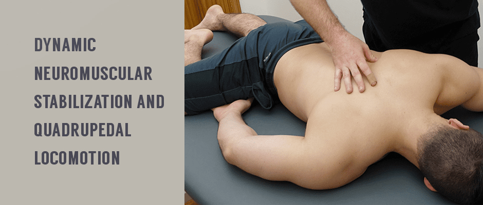
DNS (dynamic neuromuscular stabilization) is a manually assisted functional exercise method based on the developmental stages of an infant during the first year of motor development. Every infant follows specific gravity-dependent patterns as they progress by small increments, from lifting the head in a prone position to gradually walking and running. As each new skill emerges, the baby experiments and practices with it until it is mastered.
Developmental skills mastery is dependent on the maturation of the central nervous system (CNS), which governs physical maturation and is thereby responsible for the structural development of muscles, bones, and connective tissues. As the brain develops, skills mastery of motor patterns progresses, and structural development ensues.
The DNS method follows these same principles, tapping into the patient’s innate instinctual motor patterns to optimize movement. Bad postural habits, injuries and compensation patterns can cause the muscles to “forget” optimal motor patterns. The role of the therapist is to choose the right position to restore the brain-muscle connection, and to manually guide the patient into correct movement with a specific DNS skill.
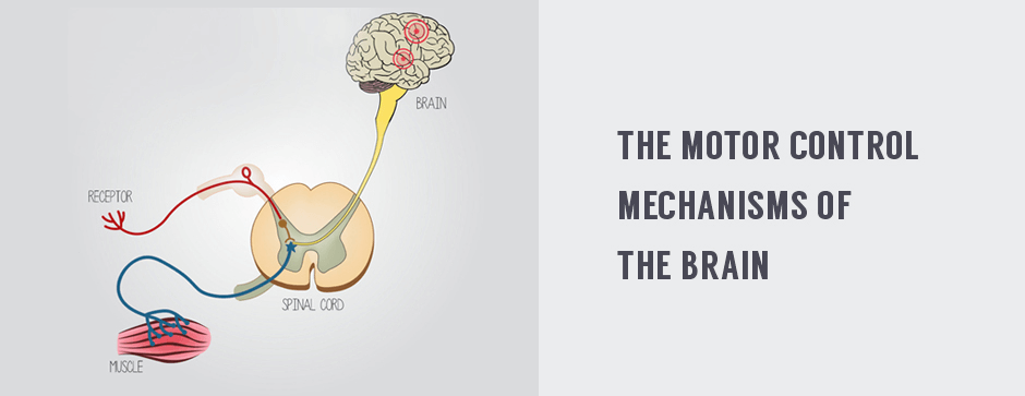
Spinal stability is key to efficient movement. Achieving spinal stability requires a 3-way interaction between the CNS, muscles and skeletal system. Conventional injury rehab and pain treatment typically focus on healing and strengthening the musculoskeletal system, to the neglect of the motor control mechanisms of the brain and central nervous system, but all three must be addressed if optimal movement is to be restored.
Intra abdominal pressure (IAP) plays a key role in spinal stability. During the early stages of an infant’s development, the diaphragm functions as mainly a respiratory muscle, but as breathing becomes coordinated with abdominal activity at around the age of 6 months, the diaphragm assumes dual roles as both a respiratory and a postural muscle. During physical activity, the diaphragm plays a critical role in stabilizing the spine during complex motor sequences.
One key posture and skill level achieved at around 9 months is quadrupedal locomotion, or crawling, where the infant learns to coordinate the movement of all four appendages to accomplish forward momentum. Crawling requires not only strength and coordination of the arm and leg muscles, but also shoulder and scapular centration, stabilization of the thoracic spine, recruitment of the core abdominal muscles, and intra-abdominal pressure to stabilize and unload the spine.

In the quadruped position, a stable spine depends on hip and shoulder/scapular centration to optimize axial extension. In this position, the core musculature is automatically activated and communicates with the contralateral oblique chains to coordinate movement.
DNS uses the crawling position to retrain the brain to properly align and stabilize the spine, to restore optimal movement. By repeating this fundamental developmental exercise, the clinician guides the patient to “remind” the muscles of their ideal motor patterns, and restore optimal connection between the CNS and the musculoskeletal system.
DNS in the quadruped position can be used to treat multiple pain syndromes, including:
Integration of ideal stabilization patterns in sports activities can potentially reduce the risk of injuries and secondary pain syndromes caused by overloading of the muscles and joints, and is likely to enhance athletic performance.
Dynamic neuromuscular stabilization is one of many innovative treatment methods we apply at NYDNRehab to help our patients heal, rehabilitate, eliminate pain and optimize movement. When used in combination with physical therapy and chiropractic care, DNS offers a simple but effective method for restoring the brain-body connection.
NYDNRehab now offers TeleHealth services, so you can get the same great physical therapy and chiropractic treatment from your home, office, hotel room, or anywhere else in the world you happen to be. Contact us today to learn more.
Resources
Frank, Clare, Alena Kobesova, and Pavel Kolar. “Dynamic neuromuscular stabilization & sports rehabilitation.” International journal of sports physical therapy 8.1 (2013): 62.

Verified Expert Profiles
Dr. Lev Kalika is a world-recognized expert in musculoskeletal medicine. with 20+ years of clinical experience in diagnostic musculoskeletal ultrasonography, rehabilitative sports medicine and conservative orthopedics. In addition to operating his clinical practice in Manhattan, he regularly publishes peer-reviewed research on ultrasound-guided therapies and procedures. He serves as a peer reviewer for Springer Nature.
Dr. Kalika is an esteemed member of multiple professional organizations, including:
Below is a prime example of how ultrasound can take the guesswork out of diagnosis.

A bad physical therapy experience is one of the primary causes of unnecessary surgery
In this instance, an athlete was originally diagnosed with minor quadriceps muscle strain and was treated for four weeks, with unsatisfactory results. When he came to our clinic, the muscle was not healing, and the patients’ muscle tissue had already begun to atrophy.
Upon examination using MSUS, we discovered that he had a full muscle thickness tear that had been overlooked by his previous provider. To mitigate damage and promote healing, surgery should have been performed immediately after the injury occurred. Because of misdiagnosis and inappropriate treatment, the patient now has permanent damage that cannot be corrected.
The most important advantage of Ultrasound over MRI imaging is its ability to zero in on the symptomatic region and obtain imaging, with active participation and feedback from the patient. Using dynamic MSUS, we can see what happens when patients contract their muscles, something that cannot be done with MRI. From a diagnostic perspective, this interaction is invaluable.
Dynamic ultrasonography examination demonstrating
the full thickness tear and already occurring muscle atrophy
due to misdiagnosis and not referring the patient
to proper diagnostic workup
Demonstration of how very small muscle defect is made and revealed
to be a complete tear with muscle contraction
under diagnostic sonography (not possible with MRI)

Complete tear of rectus femoris
with large hematoma (blood)

Separation of muscle ends due to tear elicited
on dynamic sonography examination


